Background: Limited mouth opening almost always poses many difficulties not only for laryngoscopy but also for tracheal intubation. Many patients coming for oral and maxillofacial reconstructive surgeries with limited mouth opening due to pain, scars, burns or contractures involving head and neck, distorted anatomy of head, neck and oral cavity or due to previous surgeries on the oral cavity have challenging airway. In such cases the gold standard fiber optic intubation is the choice. But other alternative techniques like blind nasal intubation or awake retrograde intubation can be a very good choices at times when fiber optic bronchoscope is not available. We chose awake retrograde intubation to secure airway in our patients. We tested two different guide wires, CVC and Zebra guide wires for railroading ET tube through nasal route in these patients having limited mouth opening of less than 2cms to evaluate the ease of intubation by comparing different parameters recorded during or after intubation. Material and methods: Intubation was performed with little modifications using two different guide wires in similar scenarios. A 0.035"F central venous catheter guide wire and a 0.032"F Zebra urological guide wire were used to guide endotracheal tube nasotrachealy. 40 patients requiring oral and maxillofacial reconstructive surgeries of various types with limited mouth opening of less than 2cms with anticipated difficult laryngoscopy and intubation were chosen. Indications for the procedure included oral submucosal fibrosis, various fractures of mandible, malignancies of gingivobuccal sulcus involving retromolar trigone, temporomandibular joint ankylosis, reconstructive surgeries of face post hemimandiblectomy, internal derangement of temporomandibular joint, submandibular abscess etc. Results: Mean time taken for successful nasotracheal intubation with Zebra guide wire Mean±SD of 3.48±0.62 minutes was significantly lesser than with CVC guide wire Mean±SD of 6.58±1.21 minutes with p<0.001. Number of attempts and patient comfort were also significantly better with Zebra guide wire, p = 0.031 and p<0.001 compared to CVC guide wire respectively. Conclusion: 0.032"F Zebra guide wire is a better, safer, inexpensive alternative to 0.035"F Cvc guide for introducing or rail roading endotracheal tube through nasotracheal route during awake retrograde intubation. Our technique of awake retrograde intubation is an effective, inexpensive, safer alternative compared to other modified techniques of awake retrograde intubation without any major complications.
Keywords
Retrograde Intubation
Cvc
Zebra guide
Hemimandiblectomy
INTRODUCTION
In the advanced era of fiber optic bronchoscope and video laryngoscope, awake retrograde intubation is very rarely used technique. But this might come handy in managing anticipated difficult airway in patients with limited mouth opening when advanced equipment is not available [1-3]. Limited mouth opening can be due to intra articular or extra articular causes [4]. Retrograde intubation was originally described by Butler F.S. Cirillo A.A. in 1960. Later modified by D.J. Waters in the early 1963. First clinical use was reported in 1981. We adopted similar but a modified technique for securing airway in our patients. Even though blind nasal intubation is technically easier compared to awake retrograde intubation, loss of airway, ET tube placement in false passage, repeated and multiple attempts, bleeding and aspiration of blood can be possibilities with this technique [3-7]. Elective tracheostomy can be another alternative but has high incidence of complications [8]. Submental and submandibular approaches are least preferred due to their technical difficulties and the complications associated [9-10].
MATERIALS AND METHODS
After obtaining institutional ethical committee clearance, informed written consent to undergo airway nerve blocks, awake retrograde intubation and to publish the data, photos etc from all the patients we conducted the study. The data was collected over a period of 8 years since it is difficult to get large number of samples in short duration of time with absolute indications for awake retrograde intubations. Only adults between 18 to 80 years of age with ASA Ι and Ⅱ status were considered for this procedure. Only patients with limited mouth opening with interincisal distance of less than 2cms were considered. All patients were intubated nasotrachealy, awake under topical anesthesia and airway nerve blocks. Intubation was performed with little modifications using two different guide wires in similar scenarios. A 0.035"F central venous catheter guide wire and a 0.032"F Zebra urological guide wire were used to guide endotracheal tube. 40 patients requiring oral and maxillofacial surgeries of various types with limited mouth opening of less than 2cms with anticipated difficult laryngoscopy and intubation were chosen11. Indications for the procedure included oral submucosal fibrosis (n = 9), various fractures of mandible (n = 7) malignancies of gingivobuccal sulcus involving retromolar trigone (n = 8), temporomandibular joint ankylosis (n = 8), reconstructive surgeries of face post hemimandiblectomy (n = 3), carcinoma tongue with carcinoma involving retromolar trigone (n = 1), internal derangement of temporomandibular joint (n = 3), submandibular abscess (n = 1). Each and every patient was assessed preoperatively for comorbid conditions, airway with specific parameters. The ease, number of attempts and time taken for intubation, nares used, change of nares done with different guide wires were noted. Any complications during intubation and in postoperative period were noted and managed accordingly.
After explaining the procedure to patient, a detailed written informed consent was taken the previous day. Nil by mouth for 6 hours was kept for all patients. Tab.Ranitidine 150mg and Tab.Alprozolam 0.5mg were given orally the previous night. On the day of surgery patient were nebulized with 4% lignocaine 4 cc for 15 minutes, three drops of xylometazoline 0.1% was instilled in both the nostrils to decongest the nasal mucosa. ENT team was kept standby for emergency tracheostomy. Patients were shifted to OT and baseline vitals were noted. Monitors included ECG, NIBP, Sp02, and Etco2. A wide bore peripheral IV cannula with 16G was secured under local infiltration and ringer lactate infusion was started. Continuous O2 insufflation was given through nasal cannula at 2 litres/minute. Inj.Glycopyrollate 0.2mg IV was administered. Superior laryngeal nerve block was given with 1ml of 2% lignocaine bilaterally12. Transtracheal block was given with 2ml of 4% lignocaine under aseptic precautions13. Cricothyrotomy was done with 16 G IV cannula and the stylet was removed. Either a 0.035"F central venous guide wire or a urological zebra guide wire of 0.032"F was passed through the cannula and advanced retrograde to bring it into oral cavity. A 06 FG feeding tube with both ends open was introduced through a predetermined nostril into the oral cavity14. Both the guide wire and the feeding tube were brought out of the oral cavity with Magill forceps and the guide wire was threaded through feeding tube to bring it out of the nasal cavity. The feeding tube was removed once the guide wire was brought out of the nose. Keeping both ends of the guide wire firm, a 7.0mm cuffed PVC ET tube in females and a 7.5mm cuffed PVC ET tube in males or whichever was appropriate was introduced over the guide wire with rail roading technique. Once the resistance was encountered for further advancement of endotracheal tube, the guide wire was removed. Confirming the tracheal placement of the ET tube was done by connecting breathing circuit and with reservoir bag movements and EtCO2 tracings on the monitor. Patient were induced with intravenous Inj.Propofol 2mg/kg, Inj.Vecuronium 0.1mg/kg, Inj.Fentanyl 2mcg/kg. General anesthesia was maintained on O2, N20 50%:50%, Sevoflurane 0.4%-2% with IPPV controlled ventilation, Inj.vecuronium as and when required. At the end of surgery, residual neuromuscular block was reversed with Inj.Neostigmine 0.05mg/kg and Inj.Glycopyrollate 0.01mg/kg intravenously. After adequate neuromuscular recovery, patients were shifted to SICU with nasotracheal tube in situ on T-piece with O2 6 liters/min. Patients were given bronchodilators nebulization and steroids to reduce possible airway edema and extubated the following day.
Time taken for successful intubation was calculated from the time airway nerve blocks were given to successful endotracheal placement of ET tube. With this we noted the number of attempts taken, time taken for successful intubation, change of nares, patient’s comfort, complications, and postoperative events if any with both CVC and Zebra guide wires. The results were noted and compared to conclude on which guide wire is better to rail road and guide ET tube in awake retrograde intubation done nasotrachealy in a scenario of limited or minimal mouth opening.
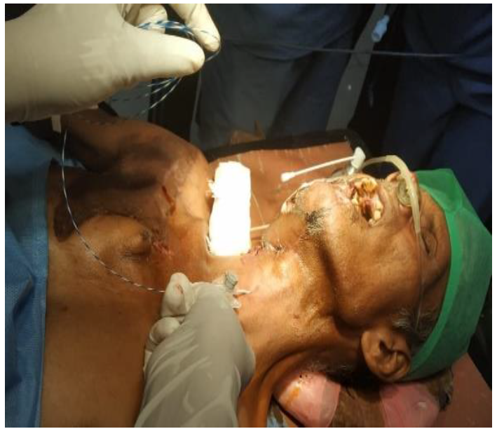
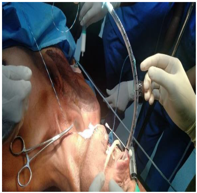
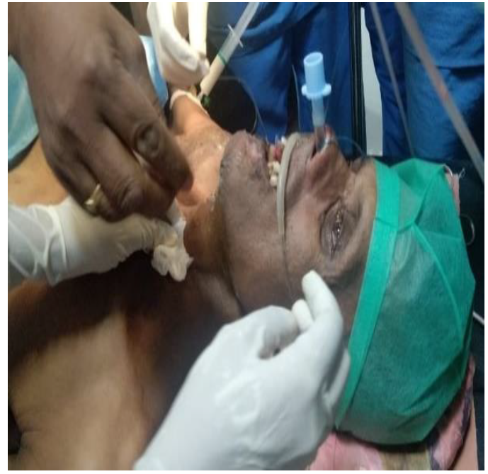
In the below paragraph we have put few photos of the procedure being performed.
Figure 1: Cricothyrotomy Done With 16G IV Cannula and Zebra Guide Wire Introduced Retrograde
Figure 2: Endotracheal Tube Railroading Over Zebra Guide Wire
Figure 3: Endotracheal Tube in Situ (Nasotracheal) After Removal of Guide Wire
The above patient is status post right hemimandiblectomy, modified radical neck dissection with an infected right deltopectoral flap. He was posted for debridement and revision of flap with reconstruction.
Statistical Methods
Descriptive and inferential statistical analysis has been carried out in the present study. Results on continuous measurements are presented on Mean±SD (Min-Max) and results on categorical measurements are presented in Number (%). Significance is assessed at 5 % level of significance. Student t test (two tailed, independent) has been used to find the significance of study parameters on continuous scale between two groups (Inter group analysis) on metric parameters. Leven`s test for homogeneity of variance has been performed to assess the homogeneity of variance. Chi-square/ Fisher Exact test has been used to find the significance of study parameters on categorical scale between two or more groups.
Significant figures
+ Suggestive significance (p-value: 0.05<p<0.10)
* Moderately significant (p-value: 0.01<p£ 0.05)
** Strongly significant (p-value: p£ 0.01)
Statistical Software
The Statistical software namely SPSS 22.0, and R environment ver.3.2.2 were used for the analysis of the data and Microsoft word and Excel have been used to generate graphs, tables etc.
RESULTS
Below we have listed few parameters which were recorded during intubation and in the post-operative period for comparison. A total of 40 patients were involved in this study with 20 patients in group Ι and 20 in group Ⅱ. In the below mentioned tables we can see that the two groups were age matched with p-0.128 with no significant differences in gender distribution p–0.337 or indications for the awake retrograde intubation. The interincisal distance in patients of both the groups were<2 cms, p–1.000. Right nare was frequently used for intubation in both the groups with 65% in group Ι, 60% in group Ⅱ without any statistical significance differences, p–0.744 but overall right nares was the most commonly used in both the groups. Changing from right to left nares for intubation was more frequent in group Ι with 20% compared to 10% in group Ⅱ but this was not of any statistical significance p–0.661. In group Ι, 55% were successfully intubated in 1st attempt, 8% in 2nd attempt and 5% in third attempt but in group Ⅱ, 90% were intubated successfully in 1st attempt and 10% in 2nd attempt without anyone requiring 3rd attempt. Thus, it’s a notable difference between group Ι and group Ⅱ, with patients very few in group Ⅱ requiring 2nd attempt, p–0.031. The mean time required for successful intubation was more in group Ι with Mean±SD of 6.58±1.21 compared to group Ⅱ Mean±SD of 3.48±0.62 requiring lesser mean time. This showed a statistically significant difference between the two groups with p-value<0.001 stating group Ⅱ required lesser mean time for successful intubation compared to group Ι. There were no complications in both the groups p–1.000 (Table 9). Patient satisfaction in group Ι were Excellent in 15%, Good in 50%, Fair in 30%, No satisfaction in 5% while group Ⅱ with Excellent in 75%, Good in 20%, Fair in 5%.
Table 1: Age Distribution of Patients Studied
Age in years
Group I
Group II
Total
<30
0(0%)
1(5%)
1(2.5%)
30-40
1(5%)
5(25%)
6(15%)
41-50
2(10%)
1(5%)
3(7.5%)
51-60
3(15%)
1(5%)
4(10%)
61-70
10(50%)
8(40%)
18(45%)
>70
4(20%)
4(20%)
8(20%)
Total
20(100%)
20(100%)
40(100%)
Mean±SD
63.25±10.16
56.55±16.35
59.90±13.86
Samples are age matched with p = 0.128, student t test
Table 2: Gender Distribution of Patients Studied
Gender
Group I
Group II
Total
Female
13(65%)
10(50%)
23(57.5%)
Male
7(35%)
10(50%)
17(42.5%)
Total
20(100%)
20(100%)
40(100%)
p = 0.337, Not Significant, Chi-Square Test
Table 3: Diagnosis / Indications for Retrograde Intubation
Diagnosis / indication for retrograde intubation
Group I
Group II
Total
Oral submucosal fibrosis
4(20%)
5(25%)
9(22.5%)
Ankylosis of TMJ
5(25%)
3(15%)
8(20%)
Ca right gingivobuccal sulcus with RMT
3(15%)
3(15%)
6(15%)
Bilateral condylar fracture mandible
1(5%)
1(5%)
2(5%)
Ca left GB sulcus with RMT
2(10%)
0(0%)
2(5%)
Internal derangement/destruction of TMJ
0(0%)
2(10%)
2(5%)
Bilateral ramus fracture
0(0%)
1(5%)
1(2.5%)
Bilateral condylar fracture mandible with left ramus fracture
0(0%)
1(5%)
1(2.5%)
Ca right lateral border tongue with gb sulcus
0(0%)
1(5%)
1(2.5%)
Internal destruction of TMJ
1(5%)
0(0%)
1(2.5%)
Left condylar fracture mandible
1(5%)
0(0%)
1(2.5%)
Left condylar fracture with right ramus fracture mandible
0(0%)
1(5%)
1(2.5%)
Multiple fracture mandible
1(5%)
0(0%)
1(2.5%)
S/p left hemimandiblectomy with PMMC flap for debridement
0(0%)
1(5%)
1(2.5%)
S/p right hemimandiblectomy with DP flap for debridement
0(0%)
1(5%)
1(2.5%)
S/p right hemimandiblectomy with pmmc flap for debridement
1(5%)
0(0%)
1(2.5%)
Submandibular abcess
1(5%)
0(0%)
1(2.5%)
Total
20(100%)
20(100%)
40(100%)
Table 4: Inter Incisor Distance- Frequency Distribution in Two Groups Studied
Inter Incisor Distance
Group I
Group II
Total
<2cms
20(100%)
20(100%)
40(100%)
>2cms
0(0%)
0(0%)
0(0%)
Total
20(100%)
20(100%)
40(100%)
p = 1.000, Not Significant, Fisher Exact Test
Table 5: Nare Used for Intubation- Frequency Distribution in Two Groups Studied
NARE used for intubation
Group I
Group II
Total
Left
7(35%)
8(40%)
15(37.5%)
Right
13(65%)
12(60%)
25(62.5%)
Total
20(100%)
20(100%)
40(100%)
p = 0.744, Not Significant, Chi-Square Test
Table 6: Change from One Nare to Another- Frequency Distribution in Two Groups Studied
Change from one NARE to another
Group I
Group II
Total
Nil
16(80%)
18(90%)
34(85%)
Right to left
4(20%)
2(10%)
6(15%)
Total
20(100%)
20(100%)
40(100%)
p = 0.661, Not Significant, Fisher Exact Test
Table 7: No. of Attempts- Frequency Distribution in Two Groups Studied
No. of Attempts
Group I
Group II
Total
1
11(55%)
18(90%)
29(72.5%)
2
8(40%)
2(10%)
10(25%)
3
1(5%)
0(0%)
1(2.5%)
Total
20(100%)
20(100%)
40(100%)
p = 0.031*, Significant, Fisher Exact Test
Table 8: Time Taken to Intubate (MINS) - Frequency Distribution in Two Groups Studied
Time taken to intubate (MINS)
Group I
Group II
Total
<5
0(0%)
18(90%)
18(45%)
5-7
12(60%)
2(10%)
4(10%)
>7
8(40%)
0(0%)
8(20%)
Total
20(100%)
20(100%)
40(100%)
Mean±SD
6.58±1.21
3.48±0.62
5.03±1.84
p<0.001**, Significant, Student t test
This clearly showed us patient satisfaction was better in group Ⅱ, p<0.001. Post-operative events were more in group Ι with 25% compared to group Ⅱ with 10% but statistically they were not significant p–0.407. Nose pain–10%, sore throat–10%, both–5% were the post-operative events in group Ι but only sore throat-10% was the post-operative event in group Ⅱ.
Table 9: Complications- Frequency Distribution in Two Groups Studied
Complications
Group I
Group II
Total
No
20(100%)
20(100%)
40(100%)
Yes
0(0%)
0(0%)
0(0%)
Total
20(100%)
20(100%)
40(100%)
p = 1.000, Significant, Fisher Exact Test
Table 10: Patient Satisfaction- Frequency Distribution in Two Groups Studied
Patient satisfaction
Group I
Group II
Total
Excellent
3(15%)
15(75%)
18(45%)
Good
10(50%)
4(20%)
14(35%)
Fair
6(30%)
1(5%)
7(17.5%)
No
1(5%)
0(0%)
1(2.5%)
Total
20(100%)
20(100%)
40(100%)
p<0.001**, Significant, Fisher Exact Test
Table 11: Post op Events- Frequency Distribution in Two Groups Studied
Post op Events
Group I
(n = 20)
Group II
(n = 20)
Total
(n = 40)
No
15(75%)
18(90%)
33(82.5%)
Yes
5(25%)
2(10%)
7(17.5%)
Nose pain
2(10%)
0(0%)
2(5%)
Nose pain, sore throat
1(5%)
0(0%)
1(2.5%)
Sore throat
2(10%)
2(10%)
4(10%)
p = 0.407, Not Significant, Fisher Exact Test
DISCUSSION
Awake retrograde intubation technique perhaps better described as wire guided blind intubation or trans-laryngeal intubation amalgamates two procedures. Retrograde insertion of a guide wire percutaneously from the larynx into mouth or nose called the “guidance” and the “blind” part is the insertion of endotracheal tube into trachea without visualization of vocal cords [8]. We included OSMF and internal derangement of TMJ [2] also as indications of retrograde intubation along with those indications mentioned by S.S. Dhara [8]. Several modifications of this technique have been made since its introduction by Butler F.S. and Cirillo A.A. [19] in both elective and emergency cases with very few complications [3-8,14-18]. In our study, we compared a “J” tipped O.O35" F CVC guide wire and a 0.032" F Zebra urological guide wire for rail roading and insertion of endotracheal tube through nasotracheal route. We used a 6 Fr gauge infant feeding tube with both open ends to bring the guide wire through nose from oral cavity in all patients since we did not want to traumatise the mucosa of nasal cavity, nasopharynx and oropharynx by repeated manipulations of guide wires while bringing them from oral cavity into the nose. In our study we brought proximal end of guide wire and distal open end of feeding tube out of the oral cavity with a Magill forceps to guide the guide wires through the feeding tube and bring them out through nose [3]. This was similar to what reported by Bhattacharya and co-workers [3] in two patients with TMJ ankylosis requiring gap arthroplasty, but used a suction catheter to retrieve an epidural catheter from the pharyngeal cavity, which had been passed retrogradely from a cricothyroid puncture to aid intubation. We did not use any guiding catheter over guide wire to intubate since the guiding catheter should be held in position firmly to prevent accidental dislodgement of tip of endotracheal tube into the oesophagus and also accidental removal or displacement of ET tube while removing the guiding catheter after endotracheal tube is advanced into final position in the trachea [2]. We removed the feeding tube before introducing the ET tube over guide wire to avoid possible difficulties in retrieval of feeding tube. We avoided overzealous pressure on the ET tube to avoid its folding [20]. Barriot and Riou [21] reported retrograde technique in 19 patients with either maxillofacial trauma or cervical spine injury and found that all were successfully intubated within 5 min in single attempt. In the present study, 55% were successfully intubated in 1st attempt, 8% in 2nd attempt and 5% in third attempt in group Ι but in group Ⅱ 90% were intubated successfully in 1st attempt and 10% in 2nd attempt without anyone requiring 3rd attempt. Thus, it is a notable difference between group Ι and group Ⅱ, with very few patients in group Ⅱ requiring 2nd attempt, p = 0.031 which is moderately significant. This showed that the 0.032" F Zebra guide wire was better than 0.035" F CVC guide wire in terms of attempts taken for successful intubations. The mean time required for successful intubation was more in group Ι with Mean±SD of 6.58±1.21 minutes compared to group Ⅱ requiring lesser mean time with Mean±SD of 3.48±0.62 minutes. This showed a strong statistically significant difference between the two groups with p-value<0.001. This implies that the time taken to successfully intubate with the Zebra guide wire was comparatively and significantly lesser than with CVC guide wire. Further this data also shows Zebra guide wire is a better guide wire significantly in terms of time taken for successful intubations. This significant differences in number of attempts and the time taken for successful intubations may be attributed to the physical nature of these two guide wires. Cvc guide wire being a vascular guide wire is even though a traumatic due to “J” tip but it is very soft, more flexible, has coiled structure without any enclosure over it which may result in expansion on stretching it or by traction on both the ends during intubation. In contrast, the Zebra urological guide wire is also a traumatic with a smaller “J” tip, soft, flexible but firm due to guide wire being enclosed with a zebra pattern silicone enclosure making it more visible, adding extra strength to it. The extra zebra pattern enclosure on Zebra guide wire makes it more firm, non-expanding on traction or stretching at both the ends while intubating. With these findings a Zebra guide wire is comparatively better than CVC guide wire for retrograde intubation in terms of number of attempts and time taken. Patient’s comfort during retrograde intubation requires effective regional anesthesia of the airway [11]. In patients with distorted anatomy, it may be difficult or almost impossible for us to perform an excellent airway nerve blocks and topical anesthesia of the airway. All thanks to our experienced anesthesiologists in performing regional anesthetic blocks of the airway. These procedures require an experienced anesthesiologist and are safe only in their hands. Patients in whom we had difficulty in manipulation or grade 2 and 3 patient comfort was not only because of tracheal tube impinging on the larynx following intubation but also due to multiple attempts taken to negotiate ET tube into the trachea as stated by Shantha T.R. [17]. In our study Patient satisfaction in group Ι were Excellent – 15%, Good – 50%, Fair – 30%, No satisfaction – 5% compared to group Ⅱ with Excellent – 75%, Good – 20%, Fair – 5%. This clearly showed us patient satisfaction was better in group Ⅱ with Zebra guide wire, p<0.001. This increased patient comfort was probably due to reduced number of repeated attempts and lesser time taken to intubate the patients with Zebra guide wire. Many retrograde and anterograde guides have been used and reviewed by S.S. Dhara [8]. Many studies have described newer and different approaches to retrograde intubation. Unfortunately some mouth opening is essential to use any of these approaches [22-25]. In case of absolutely no mouth opening, a pharyngeal catheter may be used [3]. In our study we used 16 G IV cannula to puncture the cricothyroid membrane percutaneously, a traumatic guide wires, this resulted in reduced complications as compared to other studies like Vadepally et al. [2] with complications like subcutaneous emphysema. No bleeding at the puncture site, no subcutaneous emphysema or any other complications were seen with our technique of intubation with both guide wires. Postoperatively, some patients in both the groups complained of nose pain, sore throat which resolved gradually in a day or two requiring no intervention. We did not encounter any major complications in the post-operative period also. The main advantages with our technique of retrograde intubation in patients with limited mouth opening as observed in our study were that our technique was simple, safe, effective, inexpensive, easy to perform, without any major complications and an better alternative compared to other techniques of using a guiding catheters over guide wire [2-3,8]. With all other data we also found that 0.032"F Zebra guide wire is significantly better than 0.035"F CVC guide wire with respect to number of attempts taken, time taken, and patient comfort and also with post-operative events. In patients with limited mouth opening retrieval of the guide wire through nose can be challenging but, in our study we have achieved it without any difficulty with both the guide wires we had used [3,7-8]. All these emphasized on the simplicity, safety of our modified technique of awake retrograde intubation when an expensive equipment like fiber optic bronchoscope is not available.
CONCLUSION
In our present study, our modified technique of awake retrograde intubation was successfully used in 40 patients with limited mouth opening of less than 2cms undergoing a wide variety of oral and maxillofacial surgical procedures. We conclude that a 0.032"F Zebra guide wire is the most advantageous over 0.035"F CVC guide wire for rail roading endotracheal tube during awake retrograde nasotracheal intubation in all aspects including number of attempts taken, over all time taken, patient comfort and post-operative events. Our technique also had other advantages like simplicity, minimal inexpensive equipment required, no need to identify the laryngeal inlet, no need of external laryngeal manipulation and safety of the technique in experienced hands without any complications compared to other techniques of using guiding catheter over guide wire when awake fiber optic bronchoscope is not available [8,14].
Authors Contribution: Concept and Study Design
Prashanth Gowtham Raj S.K.
Acquisition of Data, Laboratory or Clinical/Literature Search
Ethics Statement/Confirmation of Patient Permission
All patients gave informed written consent for regional anesthesia of airway, awake retrograde nasotracheal intubation and clinical images/photos for publication.
Conflict of Interest
We have no conflict of interest.
REFERENCE
American Society of Anesthesiologists Task Force on Management of the Difficult Airway. "Practice Guidelines for Management of the Difficult Airway: An Updated Report." Anesthesiology, vol. 98, 2003, pp. 1269–1277.
Vadepally, A.K. et al. “Retrograde Intubation Through Nasal Route in Patients with Limited Mouth Opening Undergoing Oral and Maxillofacial Surgery.” Journal of Oral Biology and Craniofacial Research, vol. 8, 2018, pp. 30–34.
Bhattacharya, P. et al. "Retrieval of a Retrograde Catheter Using Suction, in Patients Who Cannot Open Their Mouths." British Journal of Anaesthesia, vol. 92, 2004, pp. 888–890.
Lehman, H. et al. "Limited Mouth Opening of Unknown Cause Cured by Diagnostic Coronoidectomy: A New Clinical Entity?" British Journal of Oral and Maxillofacial Surgery, vol. 53, 2015, pp. 230–234.
Davies, J.A.H. "Blind Nasal Intubation." British Journal of Anaesthesia, vol. 47, 1975, p. 1339.
Jackson, P. et al. "Awake Blind Nasoendotracheal Intubation: A Comprehensive Review." Journal of Oral and Maxillofacial Surgery, vol. 52, 1994, pp. 1303–1311.
Gill, M. et al. "Retrograde Endotracheal Intubation: An Investigation of Indications, Complications and Patient Outcomes." American Journal of Emergency Medicine, vol. 23, 2005, pp. 123–126.
Jundt, J.S. et al. "Submental Intubation: A Literature Review." International Journal of Oral and Maxillofacial Surgery, vol. 41, 2012, pp. 46–54.
Anwer, H.M.F. et al. "Submandibular Approach for Tracheal Intubation in Patients with Pan Facial Fractures." British Journal of Anaesthesia, vol. 98, 2007, pp. 835–840.
Vadepally, A.K. et al. "Quest for an Ideal Route of Intubation for Oral and Maxillofacial Surgical Manoeuvres." Journal of Maxillofacial and Oral Surgery, vol. 15, 2016, pp. 207–216.
Simmons, S.T. and A.R. Schleich. "Airway Regional Anesthesia for Awake Fiberoptic Intubation." Regional Anesthesia and Pain Medicine, vol. 27, 2002, pp. 180–192.
Miller, R.D. Anaesthesia. Mosby Saunders: Churchill Livingstone, 2009, p. 1579.
Burbulys, D. and K. Kiai. "Retrograde Intubation." Emergency Medicine Clinics of North America, vol. 26, 2008, pp. 1029–1041.
Bourke, D. and P.R. Levesque. "Modification of Retrograde Guide for Endotracheal Intubation." Anesthesia & Analgesia, vol. 53, 1974, pp. 1013–1104.
Hines, Michael H. and J. Wayne Meredith. "Modified Retrograde Intubation Technique for Rapid Airway Access." American Journal of Surgery, vol. 159, 1990, pp. 597–599.
Shantha, T.R. "Retrograde Intubation Using the Sub-Cricoid Region." British Journal of Anaesthesia, vol. 68, 1992, pp. 109–111.
Dhara, S.S. "Retrograde Intubation—A Facilitated Approach." British Journal of Anaesthesia, vol. 69, 1992, pp. 631–633.
Butler, F.S. and A.A. Cirillo. "Retrograde Tracheal Intubation." Anesthesia & Analgesia, vol. 39, 1960, p. 333.
Wijesinghe, H.S. and J.E. Gough. "Complications of a Retrograde Intubation in a Trauma Patient." Academic Emergency Medicine, vol. 7, 2000, pp. 1267–1271.
Barriot, P. and B. Riuo. "Retrograde Technique for Tracheal Intubation in Trauma Patients." Critical Care Medicine, vol. 16, 1988, pp. 712–713.
Adebayo, Bello Seidu, Bayo Aluko Olokun, Abayomi Ademola Olaitan, and Sunday O. Ajike. "Aetiology and Presentation of Ankylosis of the Temporomandibular Joint: Report of 23 Cases from Abuja, Nigeria." British Journal of Oral and Maxillofacial Surgery, vol. 50, 2012, pp. 80–84.
Borland, L.M. et al. "Difficult Pediatric Endotracheal Intubation: A New Approach to the Retrograde Technique." Anesthesiology, vol. 55, 1981, pp. 577–578.
Mounir, N. et al. "Pulling versus Guiding: A Modification of Retrograde Guided Intubation." Canadian Journal of Anaesthesia, vol. 36, 1989, pp. 336–339.
Mahajan, R. et al. "An Alternative Technique for Retrograde Intubation (Correspondence)." Anaesthesia, vol. 56, 2001, pp. 1207–1208.
Arya, V.K. et al. "Difficult Retrograde Endotracheal Intubation: The Utility of a Pharyngeal Loop." Anesthesia & Analgesia, vol. 94, 2002, pp. 470–473.
American Society of Anesthesiologists Task Force on Management of the Difficult Airway. "Practice Guidelines for Management of the Difficult Airway: An Updated Report." Anesthesiology, vol. 118, 2013, pp. 251–270.
Recommended Articles
Research Article
Evidence Guided, Experience Based and Patient Compatible 3-Step Protocol for Managing Moderate to Severe Category of COVID19 Infections
Farah Husain,
...
Kirti N Saxena
Published: 05/01/2021
Download PDF
Research Article
A Descriptive Cross-Sectional Study to Evaluate the Knowledge about Symptoms and Risk Factors for Heart Attack (Myocardial Infarction) Among the General Population of New Delhi
Aastha Sehgal,
...
Avneet Kaur
Published: 06/05/2023
Download PDF
Research Article
Knowledge About Risk and Preventive Factors for Osteoarthritis Knee among Ortho OPD Patients in Regional Hospital, Keylong in District Lahaul and Spiti of Himachal Pradesh
Ajay Thakur,
Jaspal Chaudhary
Published: 20/06/2022
Download PDF
Research Article
A Randomized Controlled Trial Comparing the Effect of Oral Preemptive Pregabalin Compared to IV Lignocaine in Attenuating Hemodynamic Response to Laryngoscopy
P. Aashik,
...
B.N. Reddy
Published: 17/05/2023
Download PDF
PO Box 101, Nakuru
Kenya.
Email: office@iarconsortium.org
Editorial Office:
J.L Bhavan, Near Radison Blu Hotel,
Jalukbari, Guwahati-India
Raj.S.K, P. G., None, C., Amingad, B., Menon, S. L. & Kumar.K.R, V. (2021). Awake Retrograde Intubation: Our Experience and a Comparison between CVC Guide Wire and Zebra Guide Wire for Railroading. IAR Journal of Anaesthesiology and Critical Care, 2(1), 1-7.
MLA
Raj.S.K, Prashanth G., et al. "Awake Retrograde Intubation: Our Experience and a Comparison between CVC Guide Wire and Zebra Guide Wire for Railroading." IAR Journal of Anaesthesiology and Critical Care 2.1 (2021): 1-7.
Chicago
Raj.S.K, Prashanth G., Chethanananda.T.N , Bhagyashree Amingad, Sandhya L. Menon and Vasantha Kumar.K.R. "Awake Retrograde Intubation: Our Experience and a Comparison between CVC Guide Wire and Zebra Guide Wire for Railroading." IAR Journal of Anaesthesiology and Critical Care 2, no. 1 (2021): 1-7.
Harvard
Raj.S.K, P. G., None, C., Amingad, B., Menon, S. L. and Kumar.K.R, V. (2021) 'Awake Retrograde Intubation: Our Experience and a Comparison between CVC Guide Wire and Zebra Guide Wire for Railroading' IAR Journal of Anaesthesiology and Critical Care 2(1), pp. 1-7.
Vancouver
Raj.S.K PG, Chethanananda.T.N C, Amingad B, Menon SL, Kumar.K.R V. Awake Retrograde Intubation: Our Experience and a Comparison between CVC Guide Wire and Zebra Guide Wire for Railroading. IAR Journal of Anaesthesiology and Critical Care. 2021 Jan;2(1):1-7.



 Download PDF
 Download XML