Introduction: Difficult intubation carries almost one third of deaths due to failure to intubate or ventilate. We performed current study to compare tracheal intubation using Awake Fiberoptic intubation and using Intubating laryngeal mask airway in patients with difficult airway. Method-Two groups of 40 ASA I&II with MPC I,II,III were randomized for intubating trachea with awake Fiberoptic and Intubating laryngeal mask airway. Changes in hemodynamics monitored through all procedure till 10 min after intubation. Assessment done for ease of intubation,no.of attempts,complications like sore throat,arrythmias.Result-No significant difference found in demographic data as well in SBP,DBP,MAP,SPO2 in both groups. At 5 min after intubation mean heart rate changes were significant between both the groups. Maximum HR changes seen at 5 minutes as increase in HR of 18.57 from baseline in group F whereas in group I it was just 13.42.Conclusion-Intubation with Fiberoptic & Intubating laryngeal mask airway is equally successful and suitable with low complication rate in difficult airway.
Difficult intubation carries almost one third of deaths due to failure to intubate or ventilate. All patients must be examined for the ability to open the mouth and for structures visible on mouth opening, the size of the mandibular space. Traditionally, for patients with predicted to be difficult airways, establishment of a secure airway while the patient is awake is recommended. As fiberoptic bronchoscopes (FOB) have become easily available in many hospitals, awake fiberoptic intubation (AFOI) has emerged as the “gold standard” for patients with difficult airways. The term ‘AFOI’ is used to differentiates this procedure from fiberoptic intubation which is performed using proper general anesthesia. Although patients may be sedated for AFOI, but it is essential that they will remain responsive and capable of maintaining their own airway without assistance. Recently, a new device, the intubating laryngeal mask airway (ILMA) has been introduced in management of airway. It is modified version of the LMA -the ‘Intubating LMA’(ILMA) which is when combined with a modified tracheal tube can be used for blind tracheal intubation as it bypasses the pharynx and immediately allows the operator to reach the larynx. The ILMA is accepted as both rescue ventilation device and can also be used as a primary airway management device in both routine as well as in emergency departments as well as in operation theaters.
Aims and Objectives
To compare the hemodynamic changes during tracheal intubation with intubating laryngeal mask airway versus awake fiberoptic bronchoscope
After approval from institutional ethical committee,80 patients of ASA grade I and II, between 18-65 years of age, scheduled for elective surgeries under general anesthesia, were included in the study. Informed written consent in patient’s language was taken and they were divided into 2 groups of 40 patients each, by enclosed sealed envelope technique:
Group I (n = 40): Patients intubated with intubating laryngeal mask airway
Group F (n = 40): Patients intubated with fiberoptic bronchoscope
Preparation for Fiberoptic
Patients with baseline parameters noted then more potent nostril identified and IV access established.2-3 drops pf xylometazoline 0.1% instilled in to each nostril. Then 10% lignocaine sprayed over tongue and posterior pharyngeal wall and transtracheal block with 2% lignocaine given. Inj .Fentanyl 25mcg was given for sedation. Patient’s head position- sniffing of morning air position .All Tracheal intubation attempts were done nasally using a standard FOB by appropriate sized endotracheal tube(ETT).On confirmation of TI by end tidal CO2(ETCO2) detection, general anaesthesia was induced with Propofol.
Preparation for Intubating Laryngeal Mask Airway
Pt selected for intubation with intubating laryngeal mask airway were induced for general anesthesia prior to intubation. The patients received the following drugs: inj. Ondansetron 0.08mg/kg,inj.Glyco pyrrolate4mcg/kg,inj.Midazolam0.03mg/kg,inj.Fen tanyl 2mcg/kg immediately prior to induction of anaesthesia and values of HR, SBP, DBP, MAP were again noted as preinduction data. After preoxygenation with 100%oxygen for 3 minutes, induction of anaesthesia was done with inj.Propofol1–2mg/kg and inj.succinylcholine (1-2mg/kg) was given to facilitate tracheal intubation with appropriate sized endotracheal tube. In ILMA group, ILMA of appropriate size 3 or 4 ILMA was inserted and cuff is inflated 20 to 30 ml of air as checked by portex cuff pressure manometer. ILMA then attached to anaesthesia breathing system and adequate ventilation is seen by chest wall movement and capnography. When ventilation with ILMA was found to be adequate then a size of 7 or 7.5 or 8 ID well lubricated, reinforced cuffed silicone tracheal tube passed through the metal tube of intubated laryngeal mask in to trachea then cuff inflated and circuit reconnected. The correct tube placement is determined by presence of bilateral breath sounds on auscultation and also by capnography.
The HR, RR, SBP, DBP, MAP, SPO2 were recorded at baseline after induction, after intubation and after insertion of the airway device at 1,5,10 minutes. The ease of insertion of the airway device was noted.
Postoperative airway complications, including cough, blood on the device, hoarseness, sore throat trauma to tongue, lip & dental trauma, dysphonia were evaluated in the recovery postoperatively.
Paired samples t-test was used to compare parameters on the continuous scale within the group (intragroup analysis) and Student's t-test (two-tailed, independent samples) for between two groups (intergroup analysis). Results were considered significant if P value was <0.05.
The demographic characteristics (age, sex, weight) of the two groups were comparable and statistically nonsignificant.
As shown in table 1 the age of patients in two groups were comparable and there was no statistically significant difference (p<0.05)(p = 0.61)
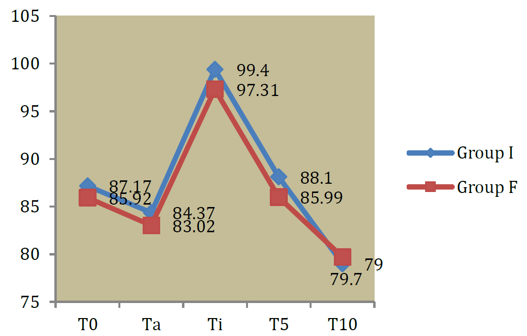
As shown in table 2 and graph 1There is no statistically significant difference in baseline values in both groups as p value is 0.07
In group I,mean baseline heart rate was 87.17±2.89 beats/min. The mean HR 5 minutes after intubation was 88.10±4.13 beats/min. The mean HR 10 minutes after intubation was 79.00±2.12 beats/min. In group F
mean baseline heart rate was 85.92±3.21 beats/min.The mean HR 5 minutes after intubation was 85.99 3.79 beats/min. The mean HR 10 minutes after intubation was 79.70±1.45 beats/min.
Table 1: Distribution of Patients According To Their Age in Years
| Age Group(yrs) | Group I | Group F |
| ≤20 yrs | 1(2.5%) | 0(0%) |
| 21-30 yrs | 12(30%) | 6(15%) |
| 31-40 yrs | 9(22.5%) | 8(20%) |
| 41-50 yrs | 9(22.5%) | 11(27.5%) |
| 51-60 yrs | 6(15%) | 11(27.5%) |
| >60 yrs | 3(7.5%) | 4(10%) |
| Total | 40(100%) | 40(100%) |
| Mean±SD | 39.73±12.98 | 43.80±12.63 |

Figure 1: Graph Showing Comparison of Heart Rate in Two Groups
Table 2: Showing the Intergroup Comparison of Mean Heart Rate (Bpm) Changes between Two Groups at Different Time Interval
| Time Interval | Group I | Group F | t-value | p-value | ||
| Mean | SD | Mean | SD | |||
| T0 | 87.17 | 2.89 | 85.92 | 3.21 | 1.82 | 0.07,NS |
| Ta | 84.37 | 3.92 | 83.02 | 3.40 | 1.64 | 0.10,NS |
| Ti | 99.40 | 4.33 | 97.31 | 3.71 | 2.31 | 0.02,S |
| T5 | 88.10 | 4.13 | 85.99 | 3.79 | 2.38 | 0.019,S |
| T10 | 79.00 | 2.12 | 79.70 | 1.45 | 1.72 | 0.08,NS |
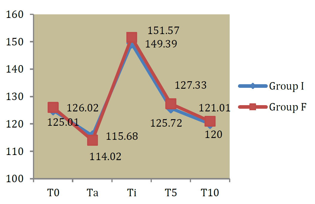
Table 3: Comparison of Mean SBP in Mmhg in Both Groups
| Time Interval | Group I | Group F | t-value | p-value | ||
| Mean | SD | Mean | SD | |||
| T0 | 125.01 | 2.89 | 126.02 | 2.21 | 1.75 | 0.08,NS |
| Ta | 115.68 | 3.96 | 114.02 | 4.11 | 1.84 | 0.06,NS |
| Ti | 149.39 | 4.84 | 151.57 | 5.06 | 1.96 | 0.05,NS |
| T5 | 125.72 | 3.64 | 127.33 | 3.79 | 1.93 | 0.05,NS |
| T10 | 120 | 1.58 | 121.01 | 3.14 | 0.23 | 0.81NS |
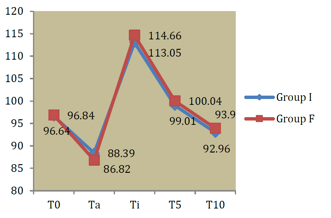
Table 4: Comparison of DBP in Mmhg in Both Groups
| Time Interval | Group I | Group F | t-value | p-value | ||
| Mean | SD | Mean | SD | |||
| T0 | 81.95 | 1.41 | 82.25 | 1.59 | 0.89 | 0.37,NS |
| Ta | 75.30 | 2.47 | 73.86 | 2.79 | 1.96 | 0.05,NS |
| Ti | 95.13 | 2.64 | 96.22 | 2.49 | 1.90 | 0.06,NS |
| T5 | 86.89 | 3.41 | 85.55 | 3.17 | 1.82 | 0.07,NS |
| T10 | 79.57 | 3.15 | 81.02 | 3.37 | 1.98 | 0.05,NS |
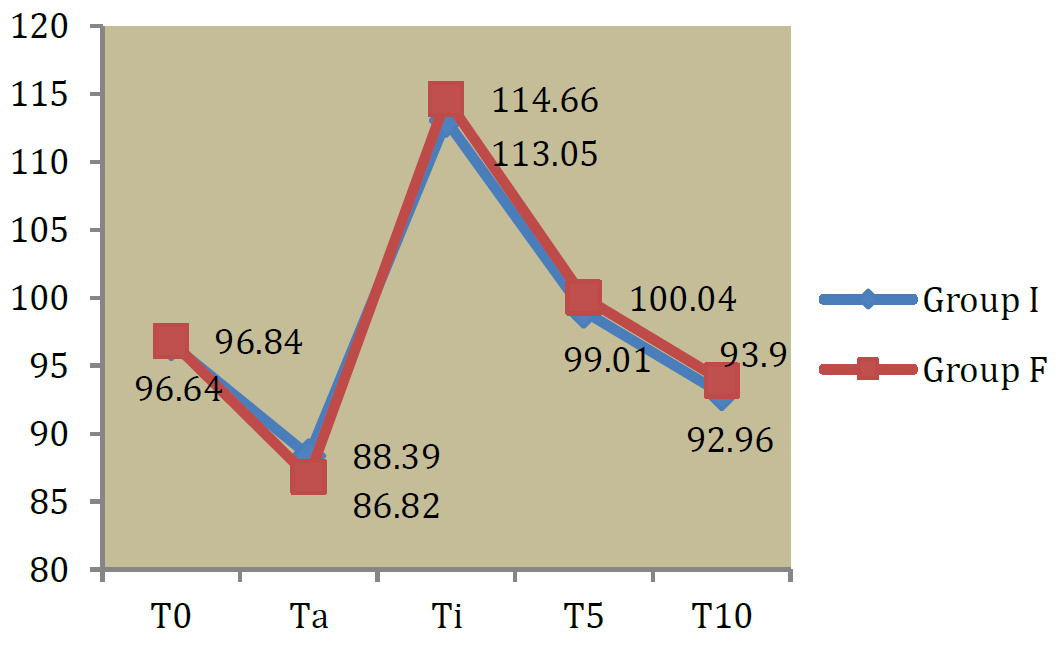
Table 5: Comparison of MAP in Mmhg in Both Groups
| Time Interval | Group I | Group F | t-value | p-value | ||
| Mean | SD | Mean | SD | |||
| T0 | 96.64 | 1.16 | 96.84 | 1.35 | 0.71 | 0.479,NS |
| Ta | 88.39 | 2.21 | 86.82 | 6.16 | 1.51 | 0.13,NS |
| Ti | 113.05 | 3.18 | 114.66 | 4.26 | 1.915 | 0.06,NS |
| T5 | 99.01 | 2.50 | 100.04 | 2.71 | 1.767 | 0.081,NS |
| T10 | 92.96 | 2.38 | 93.90 | 2.40 | 1.759 | 0.08,NS |

Figure 2: Graph Showing Comparison of SBP in MmHg

Figure 3: Comparison of DBP in Mmhg in Both Groups
Table 6: Distribution of Patients According To Total Device Insertion Time in Seconds in Two Groups
| Group | N | Mean | Std. Deviation | Std. Error Mean | t-value |
|---|---|---|---|---|---|
| Group I | 40 | 156.17 | 2.70 | 0.42 | 1.18 P = 0.24,NS |
| Group F | 40 | 159.17 | 15.83 | 2.50 |

Figure 4: Distribution of Patients According To MAP in Mmhg in Two Groups
SPO2
We found no significant difference at different time interval compared to the baseline value in both the groups.
Mostly all patients had the SPO2 in the range of 96% to 99%.
Variables and ventilation parameters of intubating laryngeal mask airway and fiberoptic bronchoscopy and revealed that there was an increase in mean heart rate after use of airway device in both groups. While basal heart rate and heart rate prior to airway placement were comparable among both groups, there was significant difference among the groups at the time of intubation and 5 minutes after intubation.
In study conducted by Hwan Joo et al, they observed that maximum heart rate during anesthetic induction were similar in both groups.
Awake fiberoptic intubation (AFOI) allows a flexible oral route or nasal route to provide visualization of vocal cords and subsequent passage of an endotracheal tube into trachea under direct vision.
Laryngoscopy and tracheal intubation can cause significant pressor responses by sympathetic stimulation. The Laryngeal Mask Airway introduced by Dr Brain in the year 1983 fulfilled to the gap between facemask and the endotracheal tube. One such modification of it is ILMA, wherein all the desirable properties of classic LMA were retained and addition it was possible to secure the airway with endotracheal tube thereby preventing the risk of hypoxia, gastric distension and aspiration pneumonitis.
Olivier Langeron et al. observed that fiberoptic intubation was successful in 45 cases (92%) and ILMA was successful in 48 cases (94%). Jadhav T et al. [1]. in their Comparative study of fiberoptic guided versus intubating laryngeal mask airway assisted awake orotracheal intubation in patients with unstable cervical spine observed that the heart rate increased in both the groups during intubation from the baseline value (p<0.001) However, there was no statistical difference between the two groups over time p = 0.38). These findings are similar to the study we conducted.
Our findings were also similar to the study conducted by Kolli S Chalam et al. who compared hemodynamic variables and ventilation parameters of intubating laryngeal mask airway and fiberoptic bronchoscopy and revealed that there was an increase in mean heart rate after use of airway device in both groups. While basal heart rate and heart rate prior to airway placement were comparable among both groups, there was significant difference among the groups at the time of intubation and 5 minutes after intubation.
In study conducted by Hwan Joo et al. [2] they observed that maximum heart rate during anesthetic induction were similar in both groups.
Conclusion
The study shows that Fiberoptic group maximum mean HR immediately after intubation was 97.31±
3.71 beats/min. (baseline: was 85.92±3.21 beats/min.) and ILMA group maximum mean HR immediately after intubation was 99.40 ± 4.33 beats/min (baseline was 87.17±2.89 beats/min.). There was no Significant difference in demographic data, SBP, DBP, MAP, SPO2 in both groups. One patient from each group had complications in the form of hypoxia. Therefore in patients with difficult airway intubation with ILMA was a successful and equally efficacious method without significant oro-pharyngolaryngeal morbidity but offers no added haemodynamic advantage compared to Awake Fiberoptic Bronchoscopy. Thus ILMA may act as a suitable alternative to Awake Fiberoptic Bronchoscopy for patients with difficult airway undergoing elective surgeries which require tracheal intubation and general anaesthesia.
Jadhav, T. et al. "Comparative study of fiberoptic guided versus intubating laryngeal mask airway assisted awake orotracheal intubation in patients with unstable cervical spine." Minerva Anestesiologica, vol. 83, no. 8, 2017, pp. 804–811.
Joo, Hwan S. et al. "The intubating laryngeal mask airway after induction of general anesthesia versus awake fiberoptic intubation in patients with difficult airways." Anesthesia & Analgesia, vol. 92, no. 5, 2001, pp. 1342–1346.
Andranik, M.D. et al. "Blood pressure and heart rate changes during awake fiberoptic nasotracheal intubation." Anesthesia & Analgesia, vol. 62, no. 10, 1983, pp. 951–954.
Langeron, O. et al. "Comparison of the intubating laryngeal mask airway with the fiberoptic intubation in anticipated difficult airway management." Anesthesiology, vol. 94, no. 6, 2001, pp. 968–972.
Lee, L.S. et al. "Clinical study of awake fiberoptic nasotracheal intubation for difficult opening mouth patients." Ma Zui Xue Za Zhi = Anaesthesiologica Sinica, vol. 28, no. 3, 1990, pp. 343–349.