
+91 6002993949
submission@iarconsortium.org
Open Access
ISSN (Print) : 2788-9459
ISSN (Online) : 2788-9467
Introduction: this study conducted to discover the protective effect of olive oil on histopathological changes that induced by administration of hydrocortisone in the kidneys. The study was accomplished in March 1, 2022– March 30, 2022 at Animal House which belonging to college of Veterinary Medicine / University of Tikrit. Material and Methods: twenty‑five male adult white mice were distributed in four cages having 5 animals each. Distilled water was administrated to the mice of 1st control Group (G1) for 10 days. The dose of hydrocortisone was injected in the femur of animals of 2nd was 1 mg per kg per day for 10 days. 3rd group (G3) animals were injected by hydrocortisone 1 mg per kg per day for 10 days and then treated with olive oil of 20 ml per day orally for another 10 days. 4th group (G4) animals were injected by hydrocortisone 1 mg per kg per day for 10 days and in the same time treated with olive oil of 20 ml per day orally. The kidneys were taken and dissected to complete the histological examination. Results: The results of the second group showed the occurrence of rare secretions at the level of the kidney tissues exposed to hydrocortisone, where fibrosis and proliferation of fibrous cells were observed, in addition to the infiltration of inflammatory cells, with the observation of bleeding and relapse of the walls of the proximal and distal urinary tubules. As for the results of the third group, the kidney tissues showed a positive response, the disappearance of most of the histological changes, and the return of the glomeruli and urinary tubules to their near-normal position, with some damage and slight degeneration in the general tissue of the kidneys, the same was the case in the fourth group, noting that the tissue damage remained more than what was in the third group. Conclusion: Through what we obtained from the results, it became clear the negative and harmful effect of the drug hydrocortisone on the kidney tissue, and in return this study proved the therapeutic and preventive role of olive oil in the treatment of tissue damage resulting from exposure to the drug.
Corticosteroids are chemical compounds that have hormonal properties and are derivatives of cholesterol. The biological efficacy of the drug is depending on the chemical structure, and due to the remarkable anti-inflammatory and immunomodulatory action of them, they usually used as a first aid in treatments of different ailments, also they maybe can act as medicine in the daily beneficiary [1].The drug (Hydrocortisone) is a fast-acting, short-acting glucocorticoid sufficient to manage adrenal gland malfunction, allergic diseases, and infections. It Cortisol has the same chemical formula and is therefore very similar to the adrenal hormones [2].
The drug is a member of the treatments that called corticosteroids, which penetrate the cell wall and binds to particular receptors in the cytoplasm to form a complex which contain the treatment in addition to the receptors, and this complex is transmitted in turn to enters the nucleus and then it binds to DNA that will be stimulated to copy a special RNA that performs the required activities in cell cytoplasm [4]. Despite the importance of hydrocortisone in the treatment of many medical conditions, it has many harmful side effects resulting from its use on the tissues and organs of the body in general [1].The Mediterranean diet is one of the most important sources of antioxidant compounds, which have been designed to be effective in preventing diabetic nephropathy [2]. Extra virgin olive oil (EVOO) is the main source of fat and at the same time an antioxidant compound in the Mediterranean region diet [3].
Beneficial effects of olive oil are specially because its high content of oleic acid. Its strong antioxidant properties are due to the presence of phenolic components. The kidney performs its vital role in the infiltration of blood and excretion of waste compounds through the various parts of its nephron [4]. It has various parts which including the Bowman’s capsule which surrounds the glomerulus. Filtration of blood takes place in the medulla through a loop of Henle and collecting tubules, both of which are found in the medulla [5].
Antioxidants such as olive oil may reduce the effect of oxidative agents on the kidney [6]. Olive oil possesses great health benefits such as the prevention of coronary heart disease, cancers, and modification in immune responses. It has anti‑apoptotic, anti‑inflammatory, and anti‑oxidative properties as it protects the tissues against damage caused by oxidative stress [7]. There is the ability of extra virgin olive oil (EVOO) to alter the cell membrane structure and to reduce oxidative injury of compounds [8]. The antioxidant effect of EVOO depends upon the amount of oleuropein and tyrosol [9]. The use of olive oil has also improved renal histoarchitecture including glomerular fragmentation, enlargement of Bowman’s space, hemorrhage, infiltration of leukocytes, and tubular dilation caused by acrylamide [10] based on its properties, olive oil may be effective in protecting kidneys from arsenic‑induced histological changes.
The drug hydrocortisone was purchased from local pharmacies and the therapeutic dose of olive oil (which obtained from Spain) was approved at the rate of 20 ml / kg of body weight, mixed daily with the diet, and given to animals (11).
In cooperation with the animal department at the College of Veterinary Medicine / University of Tikrit, the necessary animals were prepared to complete the experiment, and the experiment was completed in its affiliated departments. Twenty healthy white male mice (Mus musculus) were used in the study. The weight of the animals that were used in the experiment ranged between 30 to 35 grams, while their age was 11 to 12 weeks. These animals were divided into four groups, each group includes five animals, as follows:
The first group is control group which administerd by 1ml distilled water for 10 days
The second group which injected by 1mg/kg of hydrocortisone in thigh muscle for 10 days [1]
The third group which injected by 1mg/kg of hydrocortisone in thigh muscle for 10 days [1], and after that administered via oral dosage 20 ml/kg of oil olive with diet for 10 days [11]
The fourth group which injected by 1mg/kg of hydrocortisone in thigh muscle, and in the same time they administered via oral dosage 20 ml/kg of oil olive with diet for 10 days
Animals were starved for 24 hours to prepare them for dissection and obtaining kidneys and accomplishing of histological sections [11].
First Group
Microscopic checkup of kidneys observed normal architecture of tissues, glomerulus is surrounded by normal urinary space, and both proximal and distal convoluted tubules and normal collecting tubules were showed (Figure 1).
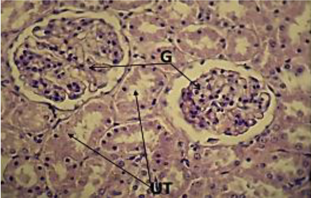
Figure 1: G1 Observes Normal Condition of Urinary Tubules (UT), and Glomerulus (G) H and E 600x
Second Group
(G2) In this group, microscopic examination of kidney tissues observed the presence of a little fibrosis, with the possibility to see a number of fibroblasts with the presence of diapedesis of inflammatory cells, also hemorrhage was visible through the renal tissues and within the lumen of many tubules, in addition to a degeneration of epithelial cells of others (Figure 2-3).
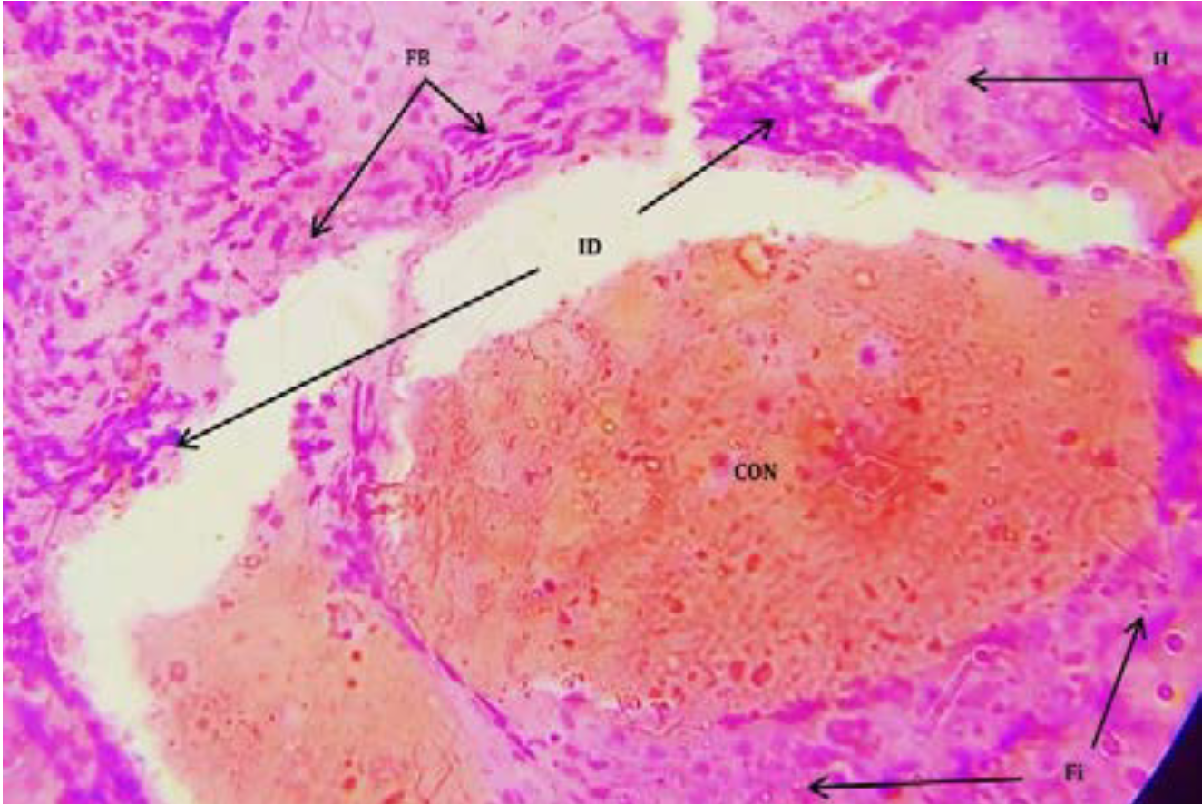
Figure 2: G2 Observes Fibroblasts (Fb), Hemorrhage (H), Fibrosis (Fi) Congestion (Con) andInflammatory Cells Infiltration (IF). H and E 400x
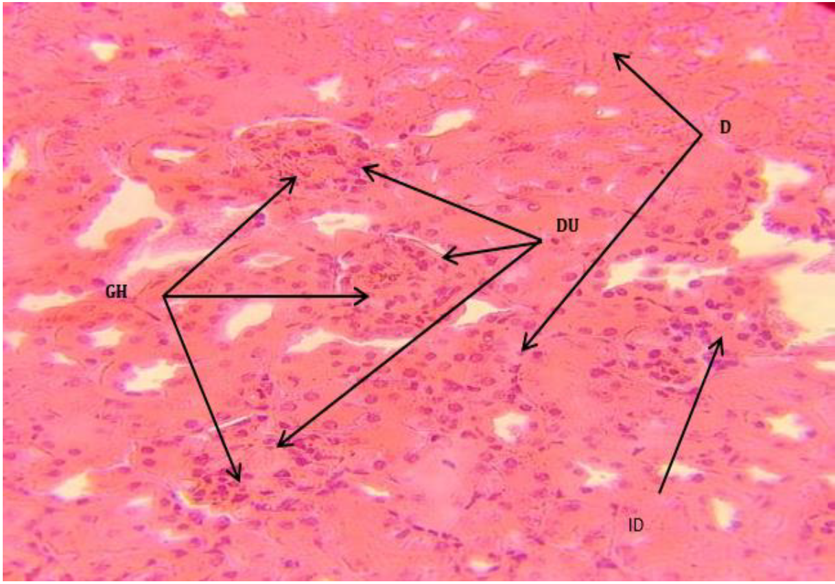
Figure 3: G2 Observes Glomerulus Hypertrophy (GH), Disappearance of Urinary Space (DU), Inflammatory Cell Diapedesis (ID) and Urinary Tubules Degeneration (D). H andE (400X)
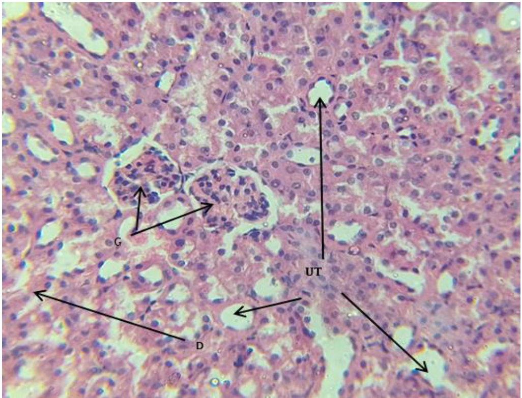
Figure 4: G3 Observes Returning of Glomeruli (G), Urinary Tubules (UT) to Normal States, and Some Degenerated Urinary Tubules (D). H and E 400x
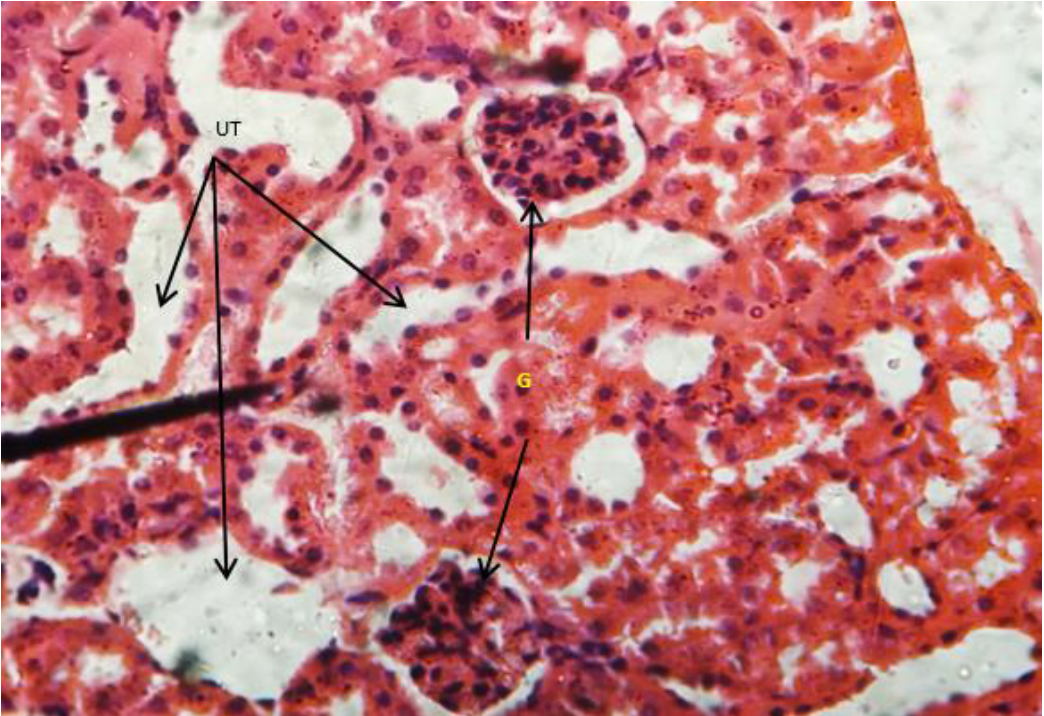
Figure 5: G3 Observes Returning of Glomeruli (G), Urinary Tubules (UT) to Normal States, and Some Degenerated Urinary Tubules (D). H And E 400x
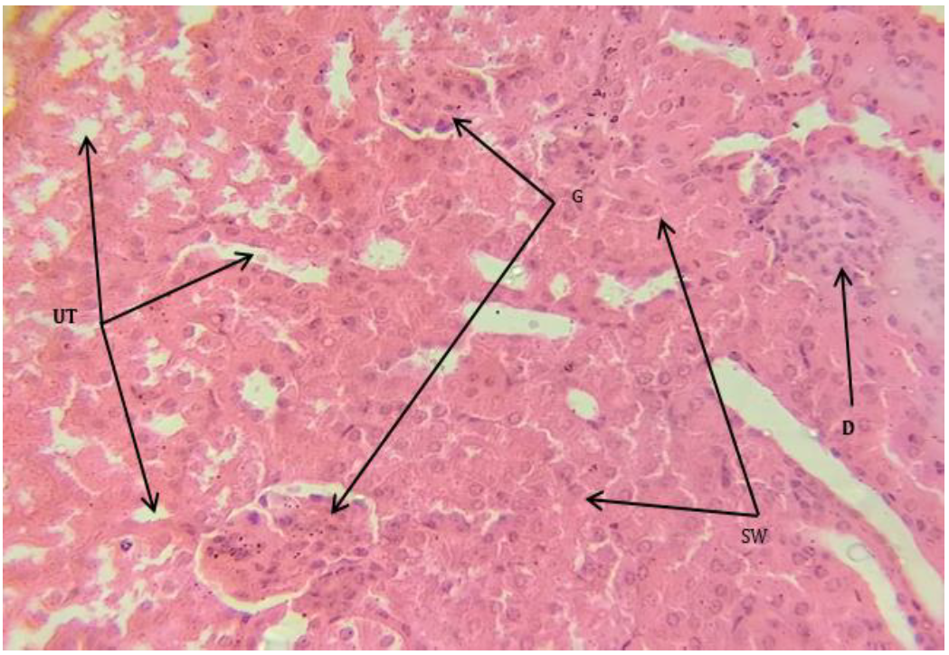
Figure 6: Observes Returning of Glomeruli (G) and Some Urinary Tubules (UT) to Normal State, with Remaining of Degeneration in Other Tubule Cells (D) with Swelling (Sw) of Others. H and E 400x
Third Group
Kidney tissues in animals of this group show a general returning to a near-nor0mal state there for glomeruli , epithelial cells that lining urinary tubules were seems to be normal and disappearance of fibrosis, fibroblasts and hemorrhage with presence of some tissue degenerations. (Figure 4-5). Fourth group: Renal tissues in animals of this group showed partially the renal tissues returned to their normal condition, as some renal glomeruli and some urinary tubes returned to their normal shape, with the possibility of cellular degeneration in the epithelial cells in some of the urinary tubes. (Figure 6-7).
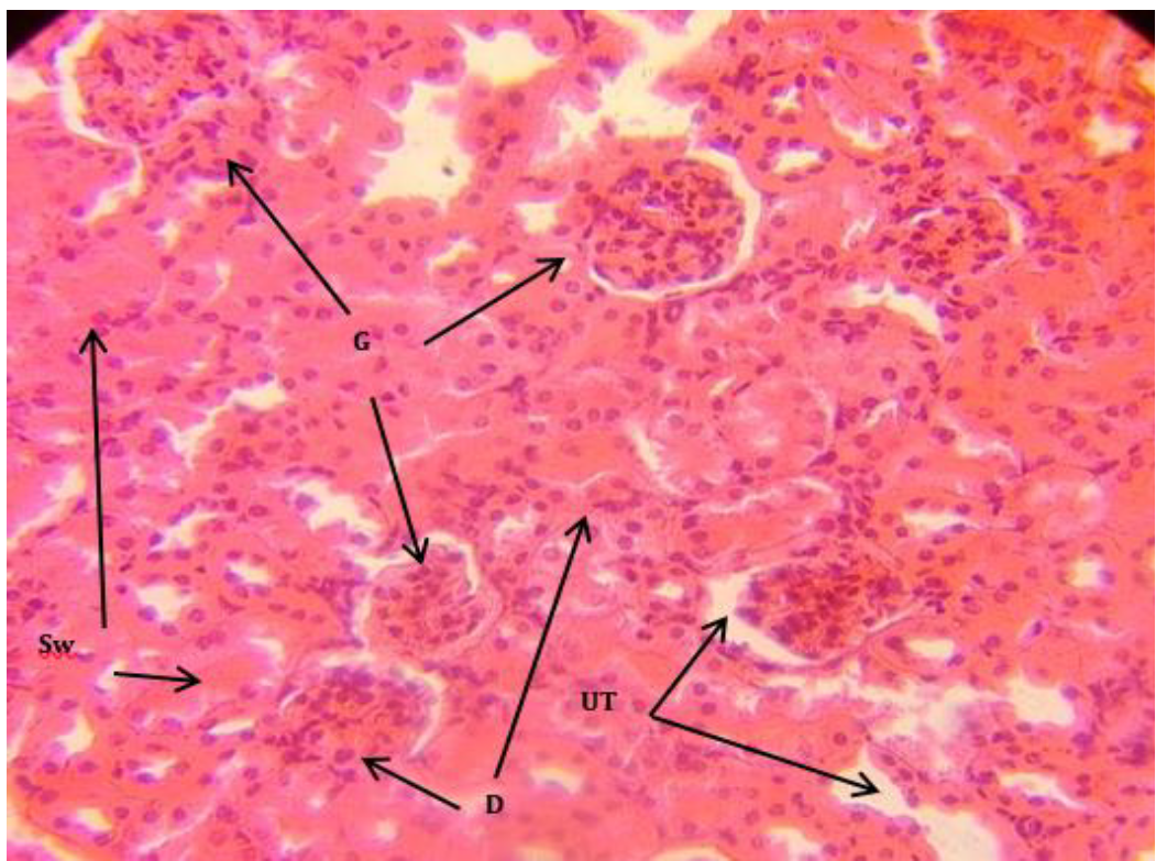
Figure 7: Observes Returning of Glomeruli (G) and Urinary Tubules (UT) to Normal State, With Remaining of Degeneration in Other Tubule Cells (D) With Swelling (Sw) of Others. H And E 400x
Microscopic examination results of the second group revale great agreement with another results of some studies, which recorded an increasing kidney weight and volume of glomeruli in mice that treated with therapeutic doses of dexamethasone ( a members of the glucocorticoid family), This represented by the presence of hypertrophy in the cells of the epithelium of the urinary tubes , as well as the presence of hemorrhage and the spreading of RBCs in kidneys tissues and a noticeable congestion of blood in the blood vessels [12]. Injury to the renal tubules as a result of poisoning leads to the death of tubular cells due to lack of oxygen, which is a critical factor in the metabolic activity of the cells of those tubules, Therefore, any damage to the blood vessels, such as necrosis or narrowing of the renal artery, leads to poor blood flow and, consequently, a lack of oxygen supply to the cells [13].
The lack of an important protein in the cell is caused by a decrease in the production of energy required for the synthesis of proteins, which are directly related to sodium pumps. Therefore, any defect in these pumps will lead to the emergence of tissue lesions and general degeneration of tissue cells, and hydrocortisone injection causes damage and necrosis of liver cells, and this leads to attracting inflammatory cells. Exposure to hydrocortisone leads to inhibition of oxidative stress enzymes in cells, and this is what will lead to damage to those cells [2].
The kidneys of the third group, G 3, were in better condition than the group In terms of response to treatment, the histological examination showed the glomeruli in their normal shape a The urinary tubules were also in good condition and very close to normal, and this is what I agree with Necib et al. [14] when they used olive oil to remove the harmful effect resulting from the act Oxidative stress caused by mercury chloride ingestion, the oxidative effect of what caused it Hydrocortisone administration was similar in terms of raising the ROS level with an increase in lipid peroxidation resulting to the formation of free radicals [15].
The repair process is due to an increase in the concentrations of the enzyme glutathione (GSH), with an increase in the effectiveness and effect of each of the enzymes GST and Px-GSH, these enzymes are the main source of anti-stress Oxidative and primary remover of the damage caused by it [11]. The positive effect of olive oil is due to It contains large quantities of tyrosol and hydroxytyrosol [16], which are natural phenols that preserve On biofilm integrity, these phenols have antioxidant properties with high affinity To sweep peroxyl radicals and the rest of the types of free radicals, as well as it has the ability to break chains of reactions producing peroxides that lead to the production of active oxygen [17]. Despite the foregoing, some damages were still found it is present in the tissue, and this may be due to the short treatment period compared to the infection period.
As for the fourth group, the preventive effect of olive oil was less than the therapeutic effect of it as it was in the third group. This may be due to the non-disappearance of the effect (hydrocortisone), but in general, the protective effect was clear in repairing the kidney tissues affected by the drug [15].
Mahdy, Sh.A. “Effect of hydrocortisone and olive oil on phagocytes.” Tikrit Journal of Pharmaceutical Sciences, vol. 13, no. 1, 2018, pp. 37–43.
Rabee, D.A. et al. “The effect of different doses from hydrocortisone on the liver tissue in the male rat.” Karbala Journal of Pharmaceutical Sciences, no. 7, 2014, pp. 58–66.
Buchman, A.I. “Side effect of corticosteroid therapy.” Clinical Gastroenterology Journal, vol. 33, no. 4, 2001, pp. 289–294.
Robles Osorio, M.L. et al. “Arsenic-mediated nephrotoxicity.” Renal Failure, vol. 37, 2015, pp. 542–547.
Schmidler, C. “Kidney anatomy and function.” Health Pages, 2022, healthpages.org.
Zhang, Q. et al. “Preventive effects of taurine and vitamin C on renal DNA damage of mice exposed to arsenic.” Journal of Environmental Health, vol. 9, 2009, pp. 169–172.
Bhattacharya, S. “Medicinal plants and natural products in amelioration of arsenic toxicity: A short review.” Pharmaceutical Biology, vol. 55, 2017, pp. 349–354.
Fitó, M. et al. “Bioavailability and antioxidant effects of olive oil phenolic compounds in humans: A review.” Annali dell’Istituto Superiore di Sanità, vol. 43, 2007, pp. 375–381.
Restuccia, D. et al. “Antioxidant properties of extra virgin olive oil from cerasuola cv olive fruit: Effect of stone removal.” Italian Journal of Food Science, vol. 23, 2011, pp. 62–71.
Ghorbel, I. et al. “Olive oil abrogates acrylamide-induced nephrotoxicity by modulating biochemical and histological changes in rats.” Renal Failure, vol. 39, 2017, pp. 236–245.
Al-Salih, M.N.M. Effect of ethanol in histopathological lesions production and biochemical varieties in liver and kidney of white rats (Rattus norvegicus) and treating by natural therapies (camel milk and olive oil) and chemical therapies (zinc acetate). PhD thesis, College of Sciences, Tikrit University, 2016.
Tayfur, S.M. Phenotypic and histological effects of dexamethasone on some organs in pregnant female white rats. Master’s thesis, College of Science, Tikrit University, 2009.
Hussein, A.J. et al. “Histopathological study of some organs after long-term treatment with dexamethasone in male rabbits.” Journal of the University of Zakho, vol. 2, no. A, 2014, pp. 39–48.
Necib, Y. et al. “Effect of virgin olive oil (Olea europaea L.) on kidney function impairment and oxidative stress induced by mercuric chloride in rats.” American Journal of Biochemistry and Biotechnology, vol. 9, no. 4, 2013, pp. 415–422.
Mustafa, M.N. et al. “Histological effect of hydrocortisone and zinc acetate in kidney of male mice.” Proceedings of the Second International and Fourth Scientific Conference, College of Science, Tikrit University, November 2020.
Mac, A. et al. “Effect of olive oils on biomarkers of oxidative DNA stress in northern and southern Europeans.” FASEB Journal, vol. 21, 2007, pp. 45–52.
Mokhtari, T. et al. “Ameliorative effect of virgin olive oil against nephrotoxicity following sub-chronic administration of ethephon in male rats.” Journal of Traditional and Complementary Medicine, vol. 10, no. 5, September 2020, pp. 487–495.
