
+91 6002993949
submission@iarconsortium.org
Open Access
ISSN (Print) : 2709-3247
ISSN (Online) : 2709-3255
Background: Tuberculosis (TB) is still a problem because children are most likely to get severe forms and it is hard to tell who has it. The goal of this paper was to describe how TB showed up in children at a tertiary level hospital, how often it happened, how it was diagnosed, and how they responded to treatment. Methods: The study looked at cases of TB in children that showed up in different ways, both inside and outside of the lungs. Polymerase chain reaction (PCR) for M. tuberculosis nucleic acids, chest X-rays, and clinical assessment were all used to diagnose the disease. All people who might have TB were given antituberculosis treatment while they waited to find out for sure. In the intensive phase of treatment, people took four drugs, and in the support phase, they took two drugs (HR). In cases where the meninges were affected, steroids were given. There were follow-up assessments. Results: In this group of cases, the symptoms of pediatric tuberculosis (TB) were different and not typical. In Case 1, a 14-year-old boy was first wrongly diagnosed with celiac disease. Later, he was found to have constrictive pericarditis and TB, which shows how important it is to think about TB when someone has a fever for a long time and other strange symptoms. In Case 2, a 2-year-old girl with TB showed up with discolored skin and at first negative PCR results. This shows how hard it is to diagnose pediatric TB. Case 3 was about a 5-year-old boy who was having neurological problems and whose brain MRI showed multiple tuberculomas. This showed how serious neurological problems can be caused by pediatric TB. In Case 4, a 2-year-old boy with stroke-like symptoms and a fever responded well to anti-TB treatment, even though the initial PCR results were negative. This shows how important it is to treat pediatric TB as soon as possible, even when diagnostic tests aren't clear. Conclusions: Pediatric TB can show up in many different and unusual ways, which makes it hard to diagnose. PCR testing was a key part of making a diagnosis. Even though the diagnosis wasn't confirmed, these cases turned out well because antitubercular treatment was started quickly. This study shows how important it is to think about TB in children who have symptoms and a history that suggest it.
Tuberculosis (TB) shows up differently in children. In 60–80% of cases, pulmonary parenchymal disease and lymphadenopathy are the most common symptoms. Extrapulmonary symptoms are also common, making up 13% of all cases. These include lymphadenopathy and problems with the central nervous system, such as meningitis, tuberculomas, and abscesses. 6 percent of cases have pleural, 5 percent have miliary, 4 percent have disseminated, and 13 percent have osteal forms [1-3]. Atypical symptoms have been seen in both children and adults, and being malnourished or having a weak immune system makes clinical diagnosis even harder. Less than half of the cases in children only affect the lungs, while the rest have mixed or only extrapulmonary involvement. Polymerase chain reaction (PCR) was used to look for Mycobacterium tuberculosis nucleic acids in different samples. Most of the positive samples came from cerebrospinal fluid [4]. Notably, the Ziehl-Neelsen stain showed that 94 percent of the positive samples were not positive. All of the children in the study had chest X-rays, which showed that 48 percent of them had macronodular patterns, 17 percent had micronodular patterns, and 13 percent had signs of consolidated pneumonia, pleural effusion, or mediastinal widening. When clinical symptoms and epidemiological factors made it seem likely that a person had TB, treatment was started before a diagnosis was made [5-6]. During the intensive treatment phase, isoniazid (H), rifampicin (R), pyrazinamide (Z), and ethambutol (E) were given together as a four-drug regimen for an average of 4.4 months. After that, the support phase had a two-drug treatment plan (HR) that took an average of 7.4 months [7-8]. Steroids were given to patients with meningeal TB, and all of them got better completely [9-11]. The study found that children under 5 years old were the most common age group for all types of TB, making up almost half of all cases [12-16]. This trend might be because people were exposed to index TB cases early on, and the BCG vaccine might protect against severe extrapulmonary TB symptoms. The average length of the study's follow-up was 4 months, which shows how important it is to keep an eye on children with TB [17]. In this study, it is important to note that three out of the five cases did not get the BCG vaccine. These cases were related to two forms outside of the lungs (miliary and meningeal) Meningeal TB is considered an emergency. Because of the risk of death and long-term effects, it is important to make a quick diagnosis and start the right treatment. In this study, signs of endocranial hypertension, cranial nerve damage, and seizures were seen in more than half of the cases of meningeal TB, along with fever. It's clear that different tests are needed to find out if someone has TB, but there is no "gold standard" test yet [7-8]. To think that a child has TB, you need to know about the history of the disease and how it shows up in the child's body. We talk about three rare cases of extrapulmonary tuberculosis that showed up in different ways.
Case 1: A 14-Year-Old Male with Constrictive Pericarditis and Tuberculosis
Patient History: A 14-year-old male appeared with a 2 month-long fever despite receiving multiple courses of antibiotics with no improvement. The patient was discovered to be underweight and to have vitiligo (a skin ailment marked by loss of skin color), ascites (abnormal fluid accumulation in the abdominal cavity), bilateral basal crepitation (abnormal lung sounds), and the absence of a BCG vaccination scar (Figure 1).
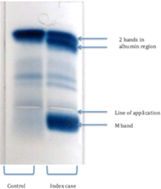
Figure 1: Case 1: A 14-Year-Old Male with Constrictive Pericarditis and Tuberculosis
Diagnostic Workup:
Various diagnostic tests were conducted:
Chest X-ray: A calcified band around the heart was discovered on a chest X-ray, raising the possibility of pericardial involvement
Echocardiography: An echocardiogram confirmed the diagnosis of constrictive pericarditis, a condition where the pericardium (the sac around the heart) becomes thickened and restricts the heart's ability to function properly
Laboratory Tests
ANA (Antinuclear Antibody): The patient had a positive ANA result, with a value of 5.3 (considered positive if >1.2). This result may suggest autoimmune activity
Blood Film: The blood film showed normochromic anisopoikilocytosis, indicating variability in the size and shape of red blood cells
ESR (Erythrocyte Sedimentation Rate): The ESR was elevated (not specified)
TSH (Thyroid-Stimulating Hormone): The TSH level was 7, which could indicate thyroid dysfunction.
Anti-ds DNA Antibody: The patient tested negative for anti-double-stranded DNA antibodies, which are often associated with autoimmune conditions like lupus
PANCA (Perinuclear Anti-Neutrophil Cytoplasmic Antibody): The PANCA test was negative, suggesting the absence of certain autoimmune disorders
Initial Diagnosis: Based on the clinical presentation and initial findings, the patient was initially diagnosed with celiac disease and placed on a gluten-free diet. However, the fever persisted
Further Investigations: Blood and urine samples were sent for culture and sensitivity testing, and double antibiotics were initiated. Despite these efforts, the patient's temperature did not subside
Tuberculosis Suspected: Given the persistent fever, imaging findings, and lack of response to other treatments, tuberculosis was suspected. Polymerase chain reaction (PCR) tests were performed on sputum and ascitic fluid, but both yielded negative results
Case 2: A Two-Year-Old Girl with Tuberculosis and Unusual Symptoms
Patient History: A two-year-old girl presented with a history of persistent cough and fever lasting for one month. Additionally, she had recently developed dark skin discoloration on the tips of her toes. Notably, the patient had a positive contact history with someone who had tuberculosis (TB)
Clinical Examination: Upon examination, the patient's blood tests revealed an elevated erythrocyte sedimentation rate (ESR) of 100 mm/hr. The blood film showed normochromic and normocytic red blood cells, and the bleeding profile was normal
Neurological Symptoms: During her hospitalization, the patient experienced repeated seizures (fits) and had daily spikes of fever
Diagnostic Imaging: A brain magnetic resonance imaging (MRI) scan was performed, revealing the presence of multiple tuberculomas in the brain with ring enhancement. This finding indicated central nervous system involvement by tuberculosis
Diagnostic Tests: A polymerase chain reaction (PCR) study on gastric lavage, performed in the early morning, initially returned a negative result for tuberculosis
Treatment: Despite the negative PCR result, given the clinical presentation and the findings on brain MRI, the patient was started on anti-tuberculosis medication. After two weeks of treatment, there was a significant and positive response to the medication, including the resolution of neurological symptoms. Interestingly, the skin discoloration on her toes also began to improve, suggesting a revascularization of affected areas
Case 3: A 5-Year-Old Boy with Tuberculomas and Neurological Symptoms
Patient History: A 5-year-old boy presented with a history of repeated fits (seizures) and a high-grade fever lasting for a few days. His symptoms were preceded by a prolonged period of lethargy, somnolence (sleepiness), and behavioral changes that had been ongoing for several months. Importantly, there was a positive contact history with a tuberculosis (TB) patient. The patient lived in a rural area, had not been vaccinated, and was exposed to a less hygienic environment
Diagnostic Workup:
To investigate the neurological symptoms and fever, the following diagnostic steps were taken:
Brain MRI: A brain magnetic resonance imaging (MRI) was performed, revealing multiple ring enhancements in the brain. These ring enhancements are suggestive of tuberculomas, which are localized, rounded lesions in the brain caused by TB infection
Tuberculosis Diagnosis: Based on the brain MRI findings, clinical history, and the positive contact history with a TB patient, a diagnosis of central nervous system tuberculosis, specifically tuberculomas, was made
Treatment: Treatment for tuberculosis was initiated promptly after the diagnosis was confirmed. Anti-tuberculosis medications were prescribed to treat the underlying TB infection
Clinical Improvement: Following the initiation of anti-tuberculosis treatment, the patient showed improvement in his condition during the next visit, which occurred one month after the treatment started. However, the patient remained on anti-TB medication as part of the standard treatment regimen
This case highlights the serious and potentially life-threatening neurological complications that can arise from tuberculosis, especially in pediatric patients. The presence of tuberculomas in the brain, as evidenced by the ring enhancements on MRI, underscores the need for early diagnosis and treatment to prevent further neurological damage and improve the patient's outcome. It also serves as a reminder of the importance of considering tuberculosis as a potential cause of neurological symptoms in individuals with relevant risk factors and clinical presentations.
Case 4: A 2-Year-Old Boy with Stroke-like Symptoms and Fever
Patient History: A 2-year-old boy was brought to the hospital with sudden-onset stroke-like symptoms, including high-grade fever and irritability. On examination, the boy had a bulging fontanelle, a noticeable swelling or protrusion of the soft spot on his head. Given the concerning symptoms and signs, a diagnostic workup was initiated.
Diagnostic Workup
Brain CT Scan: In response to the patient's symptoms, a brain computed tomography (CT) scan was performed. The results of the scan revealed temporal enhancement, indicating abnormal changes in the temporal region of the brain
Cerebrospinal Fluid (CSF) Analysis: Due to the neurological symptoms and to further investigate the underlying cause, a sample of cerebrospinal fluid (CSF) was obtained. The CSF analysis showed the presence of 200 cells, with 100% of these cells being lymphocytes. Elevated lymphocyte levels in the CSF can indicate an inflammatory or infectious process involving the central nervous system
Polymerase Chain Reaction (PCR) Test: The CSF sample was also subjected to a polymerase chain reaction (PCR) test to detect the presence of tuberculosis (TB) DNA. Despite the negative PCR result, the patient's condition did not respond to empirical treatment for meningitis
Tuberculosis Diagnosis and Treatment: Considering the clinical presentation, elevated lymphocyte count in CSF, and the possibility of TB involvement, a diagnosis of central nervous system tuberculosis was suspected. As the patient did not respond to initial empiric treatment for meningitis, anti-tuberculosis therapy was initiated
Clinical Improvement: Thankfully, a few weeks after the initiation of anti-TB treatment, the patient's condition began to improve. This positive response to anti-TB therapy indicated that the TB infection was likely the underlying cause of the neurological symptoms and fever.
This case underscores the importance of considering tuberculosis as a potential cause of neurological symptoms, especially in young children who may not always present with typical TB symptoms. It also demonstrates the value of initiating appropriate treatment, even when diagnostic tests, such as PCR, initially return negative results, as early intervention can significantly impact patient outcomes.
These four juvenile tuberculosis (TB) examples demonstrate the variety of clinical manifestations and diagnostic problems associated with TB in children. The discussion will focus on critical elements of these cases, such as the difficulty of diagnosis, the relevance of incorporating tuberculosis in the differential diagnosis, and the efficacy of anti-TB treatment. The first instance demonstrates the diagnostic difficulty posed by tuberculosis, particularly when it presents with unusual symptoms. The patient's persistent fever, vitiligo, ascites, and basal crepitation first led to the diagnosis of celiac disease, emphasizing the importance of being cautious when contemplating tuberculosis in youngsters with significant risk factors. The presence of a calcified pericardial band on a chest X-ray was a critical diagnostic finding that led to the diagnosis of constrictive pericarditis. Even when the initial presentation is unusual, this example underscores the significance of a multidisciplinary approach that includes clinical evaluation, imaging, and pertinent laboratory investigations. Furthermore, it emphasizes the importance of empiric anti-TB medication in cases where clinical suspicion is strong, because waiting for definite test findings can cause intervention to be delayed. A research on juvenile patients with extrapulmonary tuberculosis by Seo et al. [10] highlights the variety of clinical manifestations. They discovered that lymphadenitis was the most common extrapulmonary symptom in their case series, followed by pleural effusion and meningitis. This variation in presentation emphasizes the significance of keeping a high index of suspicion for tuberculosis, even when the symptoms do not match those of classic pulmonary tuberculosis. [11]
The second case, involving a two-year-old girl with tuberculosis (TB) who came with odd symptoms such as dark skin discoloration on her toes, highlights the variety of clinical presentations associated with juvenile TB. TB is known to cause a wide range of symptoms, making diagnosis difficult, especially in young children. A study by Marais et al. emphasizes the need of considering TB in patients with a history of TB contact [17]. The study stresses the significance of contact tracing in pediatric tuberculosis cases, as exposure to an infected TB case is a significant risk factor. Even if symptoms do not suggest tuberculosis right away, a history of contact should raise suspicion and require further evaluation. This is consistent with the second case, in which the patient had a positive TB contact history, which ultimately aided in the diagnosis. A study by Zar et al. supports the value of therapy trials in cases with a high clinical suspicion of tuberculosis despite initial negative diagnostic tests [18]. The paper highlights the difficulties of diagnosing tuberculosis in children, particularly when bacteriological confirmation is unclear. It implies that a favorable clinical response to anti-TB therapy can be used as a diagnostic tool when other tests are inconclusive. In the second case, the patient's favorable reaction to anti-TB medication confirms the diagnosis and emphasizes the necessity of not relying simply on negative diagnostic tests when clinical suspicion is high.
The third example, involving a 5-year-old kid with tuberculomas and neurological symptoms, emphasizes the serious neurological problems that tuberculosis (TB) can cause in juvenile patients. The ability of tuberculosis to impact numerous organ systems, including the central nervous system, emphasizes the significance of early detection and action to prevent additional neurological damage.
The existence of tuberculomas in the brain, as indicated by MRI, is consistent with the findings presented in Kumar et al. study. 's [19]. The study stresses that tuberculomas are spherical, localized brain lesions induced by tuberculosis infection. It also emphasizes that these injuries can have a variety of clinical manifestations, such as seizures, altered mental status, and focal neurological abnormalities, as demonstrated in the third example. This emphasizes the need of doctors considering tuberculosis (TB) in the differential diagnosis of pediatric patients with neurological symptoms who have a relevant history of TB exposure.
The World Health Organization (WHO) guidelines on TB management emphasize the significance of early diagnosis and fast beginning of anti-TB treatment to prevent additional neurological impairment. These guidelines emphasize the need of treating extrapulmonary tuberculosis, especially when it reaches crucial locations such as the central nervous system. Delayed treatment can lead to increased morbidity and mortality, making early intervention critical in cases like the third one [20].
The fourth instance, involving a two-year-old boy with stroke-like symptoms and fever, highlights the diagnostic problems associated with juvenile tuberculosis (TB), particularly when symptoms resemble other illnesses. Given the limitations of diagnostic testing in pediatric TB, this example underlines the importance of sound clinical judgment.
The boy's symptoms, which include stroke-like symptoms, fever, and a bulging fontanelle, are easily confused with those of other diseases. The initial negative polymerase chain reaction (PCR) results for tuberculosis (TB) may have raised initial doubts about the diagnosis. The presence of temporal enhancement on the brain CT scan and lymphocytic pleocytosis in the cerebrospinal fluid (CSF) analysis, on the other hand, elevated the possibility of central nervous system TB. These findings are comparable with those discussed in a research by Bernaerts et al. [21] that highlighted the efficacy of MRI and CSF analysis in detecting tuberculous meningitis, a form of central nervous system TB.
This example demonstrates how, in pediatric tuberculosis, clinical judgment and a strong index of suspicion can be crucial in making a fast diagnosis. It underlines that in the context of suggestive clinical and radiological symptoms, negative first diagnostic test results should not prevent doctors from evaluating TB.
The study undertaken by Marais et al. supports the need of initiating anti-TB medication early, especially in the lack of definite diagnostic tests [22]. The study found that a delay in diagnosing TB meningitis is associated with poor results, emphasizing the importance of early treatment in improving patient outcomes.
These cases highlight numerous crucial factors in the diagnosis and therapy of pediatric tuberculosis. TB can appear with a wide range of clinical manifestations, and physicians should retain a high index of suspicion, especially in high-TB-burden areas and when there is a history of TB exposure. In cases when clinical suspicion is high, the usefulness of empirical anti-TB treatment cannot be overstated, since it can lead to rapid improvement even when initial diagnostic tests are equivocal or negative. These cases serve as important reminders of the complexities of tuberculosis diagnosis and the importance of a comprehensive strategy to pediatric tuberculosis management.
Dheda K. et al. “Tuberculosis.” Lancet, vol. 387, 2016, pp. 1211–1226.
World Health Organization. Global tuberculosis report 2016.
Donald P. et al. A research agenda for childhood tuberculosis: Improving the management of childhood tuberculosis within national tuberculosis programmes—research priorities based on a literature review. WHO, 2007.
Marais B.J. et al. “The burden of childhood tuberculosis and the accuracy of community-based surveillance data.” International Journal of Tuberculosis and Lung Disease, vol. 10, 2006, pp. 259–263.
Chintu C. et al. “Lung diseases at necropsy in African children dying from respiratory illnesses: A descriptive necropsy study.” Lancet, vol. 360, 2002, pp. 985–990.
Marais B.J. and H.S. Schaaf. “Childhood tuberculosis: An emerging and previously neglected problem.” Infectious Disease Clinics of North America, vol. 24, 2010, pp. 727–749.
Secretaría de Salud, Centro Nacional de Programas Preventivos y Control de Enfermedades (CENAPRECE). Cifras oficiales de tuberculosis.
Secretaría de Salud, Dirección General de Epidemiología. Distribución de nuevos casos de enfermedad por grupos de edad en 2015. Accessed September 2016.
Nelson L.J. and C.D. Wells. “Global epidemiology of childhood tuberculosis.” International Journal of Tuberculosis and Lung Disease, vol. 8, 2004, pp. 636–647.
Seo Y.S. et al. “Xpert MTB/RIF assay for diagnosis of extrapulmonary tuberculosis in children: A systematic review and meta-analysis.” BMC Infectious Diseases, vol. 20, 2020.
Schaaf H.S. and B.J. Marais. “Management of multidrug-resistant tuberculosis in children: A survival guide for paediatricians.” Paediatric Respiratory Reviews, vol. 12, Mar. 2011, pp. 31–38.
Maltezou H., P. Spyridis, and D. Kafetzis. “Extra-pulmonary tuberculosis in children.” Archives of Disease in Childhood, vol. 83, 2000, pp. 342–346.
Sreeramareddy C.T. et al. “Comparison of pulmonary and extrapulmonary tuberculosis in Nepal: A hospital-based retrospective study.” BMC Infectious Diseases, vol. 8, 2008.
Marais B.J. et al. “Diagnostic and management challenges for childhood tuberculosis in the era of HIV.” Journal of Infectious Diseases, vol. 196, 2007, pp. S76–S85.
Verver S. et al. “Proportion of tuberculosis transmission that takes place in households in a high-incidence area.” Lancet, vol. 363, 2004, pp. 212–214.
Starke J.R. “Resurgence of tuberculosis in children.” Pediatric Pulmonology, vol. 11, 1995, pp. 16–17.
Marais B.J. et al. “A proposed radiological classification of childhood intra-thoracic tuberculosis.” Pediatric Radiology, vol. 40, 2010, pp. 816–827.
Zar H.J., et al. “Induced sputum versus gastric lavage for microbiological confirmation of pulmonary tuberculosis in infants and young children: A prospective study.” Lancet, vol. 365, 2005, pp. 130–134.
Kumar R., C.M. Pandey, and N. Bose. “Tuberculomas of the brain: A retrospective analysis of 85 consecutive cases.” Journal of Clinical Neuroscience, vol. 24, 2016, pp. 88–93.
World Health Organization. WHO treatment guidelines for drug-resistant tuberculosis (2016 update). WHO, 2017.
Bernaerts A. et al. “Tuberculosis of the central nervous system: Overview of neuroradiological findings.” European Radiology, vol. 13, 2003, pp. 1876–1890.
Marais S., et al. “Tuberculous meningitis: A uniform case definition for use in clinical research.” The Lancet Infectious Diseases, vol. 10, 2010, pp. 803–812.
