
+91 6002993949
submission@iarconsortium.org
Open Access
ISSN (Print) : XXXX-XXXX
ISSN (Online) : 2708-3594
Diseases of nails predominantly cause cosmetic disfigurement, though some may be symptomatic. Sometimes changes in nails help to corroborate diagnosis of skin disease or revealing sign of systemic diseases. Nail changes are a part of objective sign. So, these can help us in individualization, Miasmatic analysis of the patient in hand and further in Homoeopathic prescriptions. A brief review of the common and not so common nail findings in systemic diseases as well as the homoeopathic point of view of nail abnormalities.
“It’s the little things that matter the most” - Arthur Conan Doyle
Human body is a wonderful creation of God. Each and every part of the body has its own importance. Not only internal vital organs but external parts are also playing their own important role for healthy living. From these external parts, nail is one the essential parts of our human body. Whenever, our body is facing any crisis at internal organ level due to various chronic disease conditions, it also reflects its disturbance on the external part. Systemic disease typically affects more than one nails. Fingernails usually provide more accurate information than toenails, because clinical signs on toenails are often modified by trauma [1,2].
Examination of Nails
The condition of the nails may reflect both local and systemic disease. Examination of nails is important though more so in medical cases. Take a detailed history and try to find out the cause followed by a complete physical examination including dermatological, systemic, nutritional or traumatic history that may provide clues to the diagnosis of the underlining disease condition [3].
Things to observe during the examination of nails:
Pitting/non pitting
Clubbing/changes in the shape of the nails
Nail fold infraction
Splinter hemorrhage
Other lesions i.e. any changes in the surface of nails e.g., Brittle nails, onycholysis etc.
Abnormal Changes in Nails and Systemic Diseases
Abnormal Changes in Shapes
Clubbing: The finger nails have tended to bulge and are increased convex curvature in all directions and loss of the angle between nail and nail fold (Figure 1).
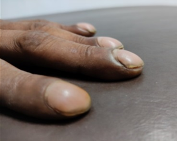
Figure 1: Clubbing
Pathogenesis of Clubbing
This condition appears due increased blood flow through vasodilated capillary plexus due to low blood oxygen level and strongly related to megakaryocytes. Any disruption to normal pulmonary circulation i.e., inflammations, cancer right to left shunt, would allow large megakaryocyte into the systemic circulation. They become lodged in the capillaries of the fingers and toes, releasing platelet- derived growth factors and vascular endothelial growth factors, which lead to tissue growth factors, vascular permeability and recruitment of inflammatory cell; this may be the probable cause.
Diagnosis:
Schamroth’s Window: It is seen when the dorsal aspects of two fingers from opposite hands are opposed, revealing a window of light, bordered laterally by the lavibond angles. As this angle is obliterated in clubbing, the window closed (Figure 2)
Lovibond’s Angle: The junction between the nail plate and the dorsal surface of the distal phalanx forms an angle, which is normally less than 160 degrees; however, this is altered to over 180 degrees in clubbing (Figure 3)
Curth’s Angle: The distal inter phalangeal joint, which is normally about 180 degree, is diminished to less than 160 degree in clubbing (Figure 3) [4]
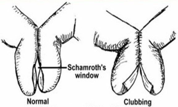
Figure 2: Schamroth’s Sign
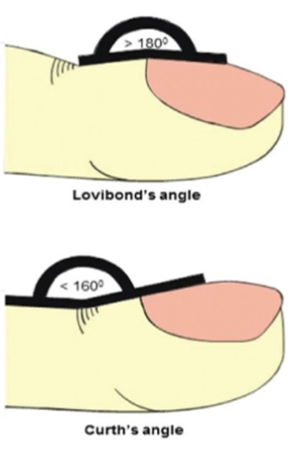
Figure 3: Lovibond’s Angle and Curth’s Angle
Cause
C-Cyanotic congenital heart disease
Cystic fibrosis
Cirrhosis of liver
Crohn’s disease
L-Lung carcinoma
Lung abscess
U-Ulcerative colitis
B-Bronchiectasis
B-Benign mesothelioma
I-Infective Endocarditis
N-Neurogenic tumor
G-Gastrointestinal disease e.g., Coeliac disease, GI lymphoma
Unilateral Clubbing
Hemiplegia
Vascular lesion-upper limb artery aneurysm, Takayasu’s Arteritis, Brachial arteriovenous malformation
Hemodialysis fistulas-iatrogenic [4]
Koilonychias
The term “koilos” in Greek means ‘Hollow’. This is a condition where the reverse or concave curvature of the nail is formed. The nail become thin and softened, and edges everted in such manner that it looks like spoon, hence also known as Spoon shaped nail. Both finger and toe nails may be affected (Figure 4).
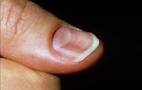
Figure 4: Spoon Shaped Nail
Cause
Iron Deficiency Anemia (most common), Plummer Vinson syndrome, Hemochromatosis, Malnutrition, Endocrine disorder e.g., Acromegaly, hypothyroidism
Platynychia/Flat Nail
The Greek word “platy” means ‘flat’ or ‘broad’. This is an abnormal condition of nail where the nail surface becomes flat and broad (Figure 5) Flat nail Suggest Iron deficiency anemia, hereditary.
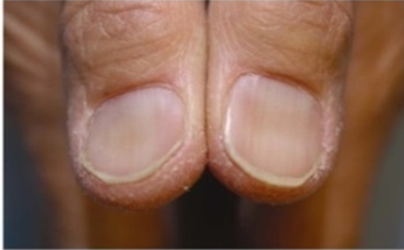
Figure 5: Flat Nail
Anonychia
It is a condition where absence of nails of finger and/ or toes (Figure 6). This condition can be associated with congenital developmental anomaly, or congenital Ectodermal defect.
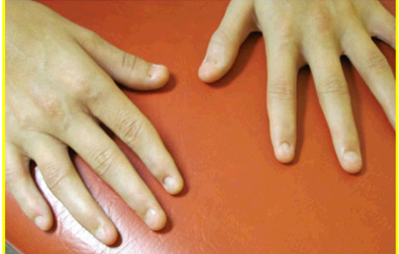
Figure 6: Anonychia
Cause
Other reasons of absence of one or more nails are severe infection, severe allergic contact dermatitis, Raynaud’s phenomenon, lichen planus.
Associated with:Onchoatrophy seen in Nail patella syndrome, congenital ectodermal defect
Missing Nails: Found in Nail patella syndrome with hereditary Nephrotic syndrome
Bitten Nails/Onchophagia
The word “phagia” Derived from the greek word ‘phegein’ meaning ‘To eat’ or ‘devour’. It an abnormal habit of consumption of finger nails by biting them (Figure 7). This type of habit commonly found in children. This habit may lead to infection of nail bed like paronychia as well as GI troubles. Suggest: Anxiety neurosis.
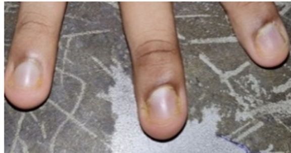
Figure 7: Onchophagia
Abnormal Nails Attachment
Onycholysis/Plummer Nails
The word “lysis” meaning ‘breakdown’ or ‘destruction’ or ‘loosening’. Onycholysis is an abnormal clinical condition where spontaneous distal separation of nail from the nail bed (Figure 8) distal detachment is commonly seen.

Figure 8: Onycholysis
Cause
Hyperthyroidism/Graves’ disease, Fungal infection e.g., Candidiasis, ringworm infection, Lichen planus, Psoriasis, trauma.
Onychomadesis
It is the proximal separation and falling off of nail plate from nail bed (Figure 9). Common cause of this condition is infection to the nail or any trauma to the matrix of nail. If the cause of disease is removed a new nail will form.
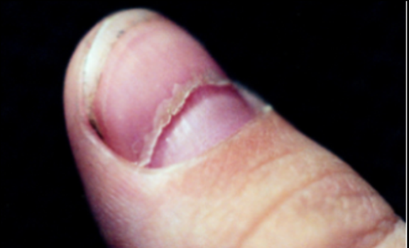
Figure 9: Onychomadesis
Cause
Trauma to the nail matrix, Pemphigus, Malnutrition, hypocalcemia, radiation.
Onychomycosis
It is fungal infection of nail. Most common causative organism responsible for the infection is ‘Trichophyton rubrum’. Here the nails become yellowish, thick, misshaped. Great toe affects more often. Other fungus which may cause onchomycosis are Candidiasis, ringworm (tinea unguium) (Figure 10).
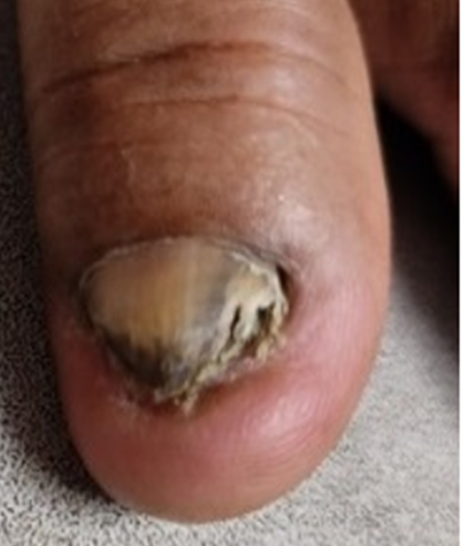
Figure 10: Tinea Unguium
Cause
Beside the infection some other cause which may leads to this condition are psoriasis (most common), Trauma, poor peripheral circulation, Pellagra.
Hangnails
This is a condition where there is splitting and peeling off cuticle of nail due to overextension of the cuticles from the proximal and lateral nail fold (Figure 11). The area becomes sensitive and painful.
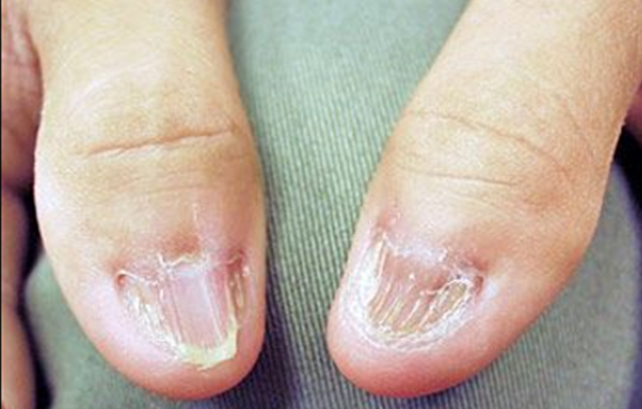
Figure 11: Hangnails
Cause
Dry skin, nail biting, using of harmful chemicals, in some cases of diabetes.
Onychogryphosis
It is a condition where the nails become thicken, yellowish opaque and claw shaped or a ram’s horn (Figure 12). It is usually due to neglect or failure to cut nails for a long time. This condition commonly found in Old age.
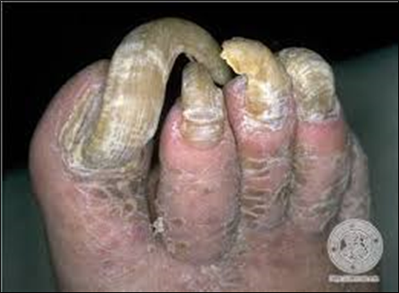
Figure 12: Ram’s Horn Nail
Onychophosis
It is a condition where thickening of nail due to diffuses hyperkeratotic tissue deposited on the lateral or proximal nail fold within the space between the nail fold and the nail plate (Figure 13). It is also seen in old age.
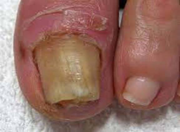
Figure 13: Onychophosis
Onychauxis
It is simply hypertrophy and thickening of nail without any deformity. Over times the nails become yellowish, hard (Figure 14).
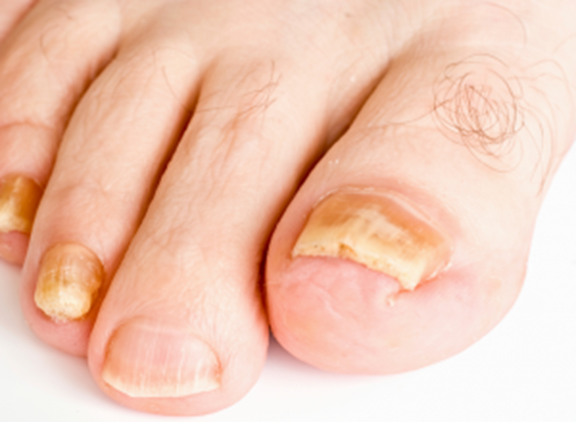
Figure 14: Onychauxis
Seen in: Acromegaly, psoriasis, Pityriasis rubra,
Pterygium Unguis
There is an inflammatory destructive process that precedes the pterygium formation, where the nail matrix is destroyed by the inflammation and replaced by fibrosis and growth of the cuticle on to the nail plate (Figure 15).
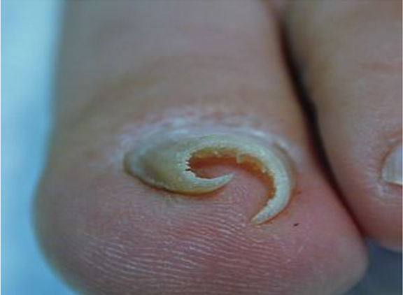
Figure 15: Pterygium Unguis
Cause
Lichen planus, Hansen’s disease, sarcoidosis, idiopathic atrophy of the nail.
Onychocryptosis
The word “cryptos” Derived from the Greek word “KRYPTOS” means ‘hidden’. It is a painful condition where the nail grows lateral side of the nail plate and it cuts the side of the nail bed causes inflammation and bleeding from the nail fold. This condition also knows as Ingrowing toe nail (Figure 16). The lateral margin of the nail acts as a foreign body and may cause exuberant granulation tissue. It is commonly seen in great toe.
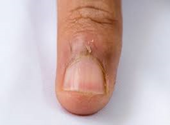
Figure 16: Ingrowing Toe Nail
Cause
Most frequently due to wearing unfitted shoe or tight shoe. It occurs sometime due to deep cutting of nail.
Abnormal Changes of Nail Surfaces
Longitudinal Striations: These are narrow furrows or grooves present the whole length of the nails. There may be associated with discoloration and thickening of the nail (Figure 17). This type of lines or ridge normally found on finger nails due to normal ageing process, hence it is very common in old age.
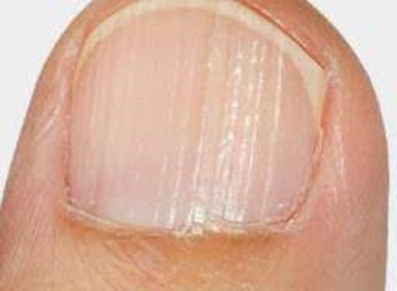
Figure 17: Longitudinal Ridge
Cause
Hereditary, history of trauma. Some other conditions that tend to cause of these grooves are lichen planus, Daries’s disease, Peripheral vascular disease, Rheumatoid arthritis.
Beau’s Lines
These are the transverse furrows from temporary arrest of nail growth at times of biological stress (Figure 18).
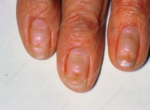
Figure 18: Beau’s Line
Cause
Severe infection, mal nutrition, diabetes.
Thimbles Nails/Pitting Nails
This condition characterized by multiple small shallow or deep depression or punctate erosion on the nail surface (Figure19).
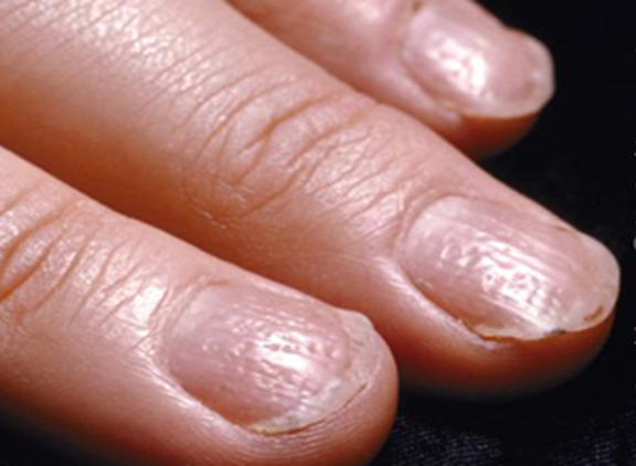
Figure 19: Pitting Nails
Cause
Psoriasis, Atopic eczema, Alopecia areata, early stage of lichen planus.
Onychoschizia
The term “Schizo” in Greek is ‘Division’ or ‘split’. It is a condition where transverse (commonly) or longitudinal splitting of nail plate into two layers near the free edge (Figure 20).
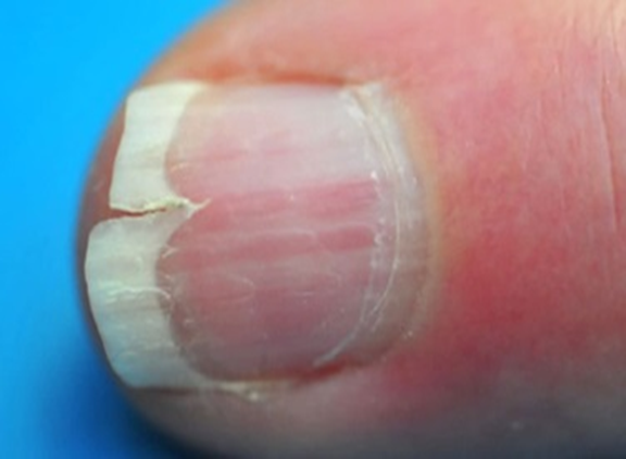
Figure 20: Split Nail
Cause
This condition occurs commonly due to Dehydration, cronkhite-Canada syndrome (multiple intestinal polyps, diarrhea, nail deformity, hair loss).
Trachynychia
“Trachy” is a greek word means ‘rough’ or ‘uneven’. So the name Trachynychia refer to the roughness of nails. It is a clinical condition where all the nails of finger and toes become dull, rough, having uniform longitudinal ridge along the nail surface (Figure 21). Sometimes this condition also known as ‘Twenty nail dystrophy’ where all the twenty nails of fingers and toes are involved.
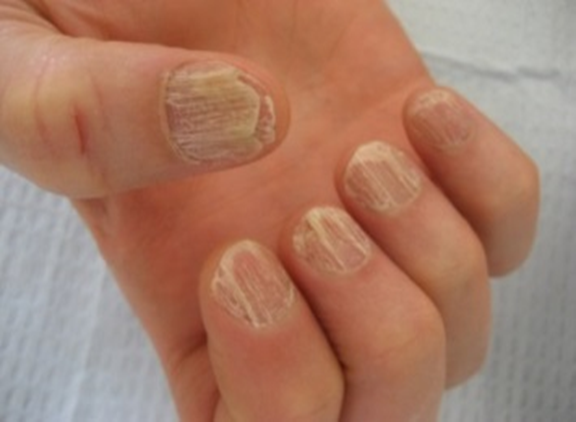
Figure 21: Trachynychia
Cause
This condition associated with Psoriasis, lichen planus, atopic dermatitis, ichthysis vulgaris. It is often seen in Vitiligo, IgA deficiency.
Onychorrhexis/Brittle Nail
The term “Rhexis” means ‘rupture, bursting, and section’. This is a clinical condition where brittleness of nails with subsequent breakage of nails of finger and toes (Figure 22).
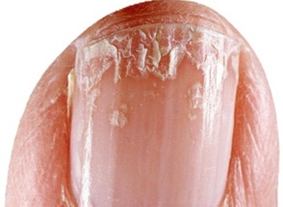
Figure 22: Brittle Nail
Cause
Use of strong soap, nail polish, fungal infection, Iron deficiency anemia, Psoriasis, zinc deficiency, biotin deficiency, Peripheral vascular disease, Hypothyroidism.
Shiny Nails
It is a condition that signify frequent rubbing of eczematous skin elsewhere.
It is an indirect evidence of pruritus, eczema.
Abnormal Changes in Color of Nails
Melanonychia: It is a condition where blackish or brownish discoloration of the normal nail plate, usually in the form of vertical or horizontal Strips along the nail plate single or multiple or in spots. This condition commonly associated with trauma to the nail, vitamin B12 deficiency.
Hutchinson Sign
It is a blackish or brownish longitudinal line along with nail plate (Figure 23), found in subungual melanoma
Also found in some systemic disease like Addison’s disease, bowen’ disease, Laugier Hunziker syndrome, Peutz- Jeghers Syndrome, subungual melanoma
Iatrogenic cause like chemotherapy, X-ray radiation, hair dyes
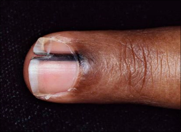
Figure 23: Hutchinson Sign
Gray Black Nail
Melanoma.
Leukonychia/White Nails
It is a condition where the changes in the nail bed are responsible for whitish appearance of nail (Figure 24).
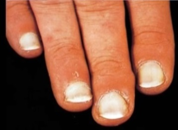
Figure 24: Leukonychia
Cause
Hypoalbuminaemic conditions like cirrhosis of liver, Nephrotic syndrome, severe malnutrition, Kwashiorkor, and Protein losing enteropathy, White spot in nails is due to Hypocalcemia. Nail bed pallor can be a nonspecific sign of severe anaemia.
Terry Nails
It is a condition where due to changes in the nail bed, the proximal portion of nail become white/pink, nail tip is red/brown (Figure 25). These changes are especially seen in patients suffering from Cirrhosis of Liver, Chronic kidney disease, Congestive cardiac failure.
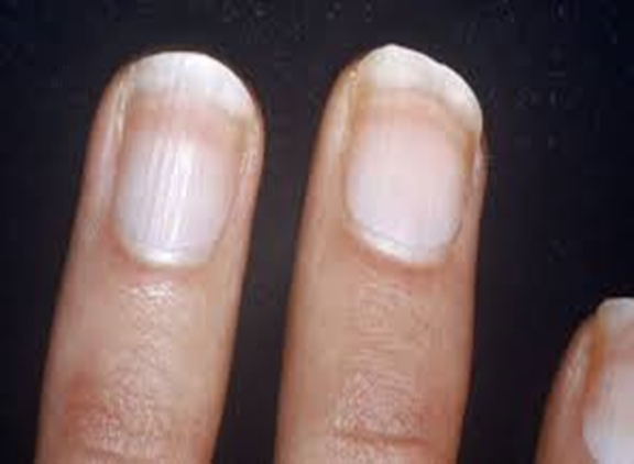
Figure 25: Terry’s Line
Mee’s Line
It is a condition where there is single white transverse band in the nail bed (Figure 26). Classically found in Arsenic Poisoning, Carbon monoxide poisoning and chronic kidney disease.

Figure 26: Mee’s Line
Muehrcke’s Band
These are paired white parallel transverse lines occur in the nail bed without furrowing of nail itself (Figure 27). A disturbance in the nail bed, where due to edema there is a slight separation of the normally adherent nail from its bed, seems to be likely cause. These lines or bands are disappearing on pressure.
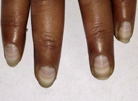
Figure 27: Muehrck’s Band
These conditions suggest chronic Hypoalbuminaemia, Hodgkin’s disease, Pellagra.
Blue Lunula (arura ares)
It is a clinical condition where there is blueish discoloration of nail especially distal part of nail bed (Figure 29).
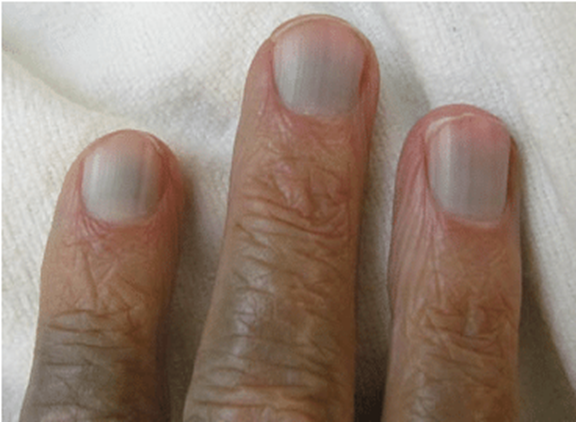
Figure 29: Blue Lunula
Cause
CuSO4 poisoning, Wilson’s disease, Cyanosis, Ochronosis
Red Lunula
It is a condition where there is dusky erythema of the lunula area, it is less inkstands in distal part of the nail where it merges with the nail bed (Figure 30).
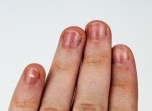
Figure 30: Red Lunula
Cause
Usually associated with Alopacia areta, history of psoriasis, Congestive cardiac failure, Chronis obstructive pulmonary disease, Reticulosarcoma
Green Nail
It is a clinical condition where there is dark green pigmentation of the nail due to exposure of nail to the bacterial organism (Figure 31). This condition is found in Green nail syndrome, caused by Peudomonas aeruginosa.

Figure 31: Green Lunula
Yellow Nail Syndromes
It is a clinical Condition where the nails become thick, and yellowish. The nails are markedly curved both transversely and longitudinally (Figure 32). The nails in this condition grows very slowly. Other components of the syndromes are lymphedema of the extremities and pleural effusion, prolonged tetracycline therapy.
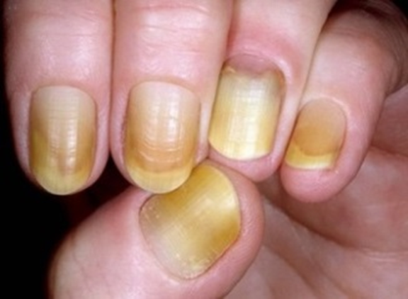
Figure 32: Yellow Nail Syndrome
Half–Half Nails/Lindsay Nail
A condition where there is proximal half of the nail is white and distal half is red or pink, with a sharp demarcation between two (Figure 28).
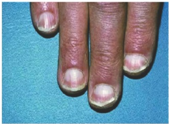
Figure 28: Half Half Nails
Seen in Uremia.
Abnormalities in Nail Bed and around the Tissue Fold:
Paronychia/Whitlow: It is infection and inflammation of the Nail fold and present as a painful swollen nail with intermittent discharge (Figure 33)
Cause: Working in wet conditions, Diabetes, Repeated trauma
Subungual Fibroma/Koenen’s Fibroma: Seen in Tuberous sclerosis (Figure 34)
Horder’s Line or Splinter Hemorrhage: These are fine longitudinal hemorrhagic streaks under the nail, which in febrile patient may suggest Infective endocarditis (Figure 35)
Cause: Sub acute endocarditis, trauma, trichinosis, systemic vasculities, vitamin c deficiency
Nail Fold Thrombi: Nail fold infract typically seen in vasculitic disorders, Scleroderma (Figure 36) [3]
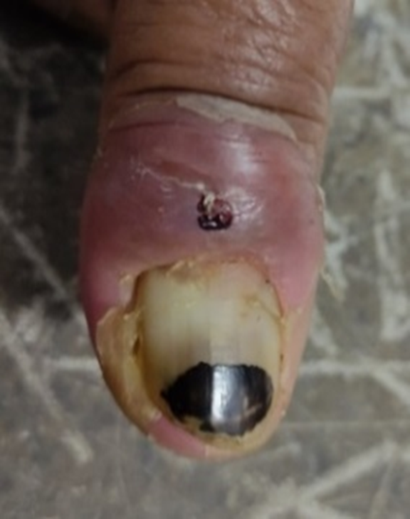
Figure 33: Paronychia
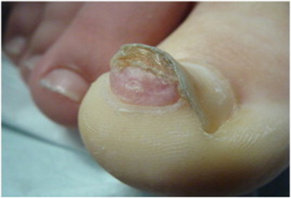
Figure 34: Subungual Fibroma
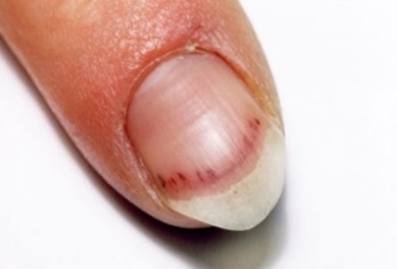
Figure 35: Splinter Hemorrhage
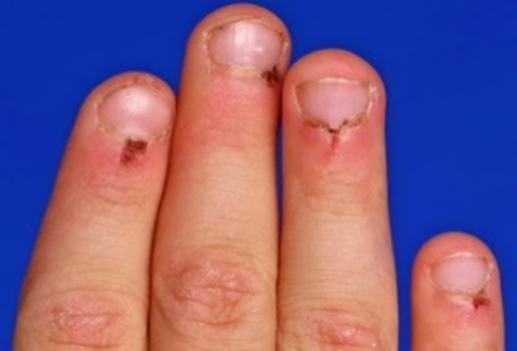
Figure 36: Nail Fold Thrombi
Miasmatic Analysis of Nails Abnormalities
Allen [5-7] (Table 1).
Table 1: Distribution of Nail Abnormalities Based on Miasmatic Analysis
Psora | Sycosis | Syphilis | Psuedo-psora |
|
|
|
|
Repertorial Approach in Nails
Kent Repertory: Different types of abnormal nails rubrics are found in Kent’s repertory under the chapter Extremities. Rubrics related to nails are found in scattered manner due to alphabetical arrangement of all the rubrics [8]:
1. Blood, oozing from finger nails – crot.h
2. Brittle, finger nails – Graph, psor
Toe nails – sil, thuj
3. Claw, like finger nails – Ars.
4. Constriction, under nails, cramps like – elaps.
5. Corrugated nails – Sil., thuj
6. Cracks, nails on – Ant.c, nat.m. sil. Ars
7. Crippled, finger nails – Graph. Sil. Caust. Nit.ac. sabad. Sep. thuj. Alum. Sulph
Toe nails – Graph. Sil.
8. Curved finger nails – Nit.ac.
9. Deformed (See distorted)
10. Detached (See separated)
11. Discoloration, finger, nails: Ant. C. Ars. Graph. Nit.ac. thuj
Black – Ars. Graph.
Blood settle under nails – apis
Blueness – nux.vom. verat
Dark – morph. Ox. Ac
Gray – merc.c. sil.
Livid – Colch. Ox.ac. Ars. Op. sul.ac
Purple –
Red – ars. Crot.c. lith.
then black - ars
white – cupr. Nit. Ac.
Spot – Sil
Yellow – Con., Sep., Sil., merc., nit-ac., nux-v., sulph.
12. Distorted nails – Graph. Sil. Fl.ac., sep. thuj alum. Anan. Calc. merc. sabad. Sulph
13. Dryness, nail about – Sil. Nat.m
14. Eruptions, nails about – Eug. Merc. Sel.
15. Exfoliation, nails – Graph
16. Felon (onychia, paronychia, panaritium), beginning in nails – Sil. Phyt. Rhus.t. sulph
17. Grow, nails do not – ant.c.
Rapid – fl.ac.
18. Hang nails – Nat.m. sulph. Cal. Merc. Rhus.t. Sil. Stann. Thuj. Lyc. Sabad. Sep.
19. Hardness, finger nail – Ars.
20. Horny, growth under nails – Ant.c. Graph
21. Inflammation, nail – Kali.c.
22. In growing toe nail – Mag.aust. Sil. Graph. Teucr. Caust. Nat.m. Nit.ac. Ph.ac. Sulph. Thuj
23. Injuries, nails, of - Led. Hyper
24. Itching, nails, around – Hep. Merc.
25. Pain, nails – Caust. Graph. Hep. Merc. Nit.ac. Nux.v. petr. Sil. Squil.
26. Pulsation, nail around – Con.
27. Split nails – Ant.c. Sil. Squil. Sulph
28. Spotted nail – Sil. Nit.ac. Alum. Ars. Ph.ac. Sep. Sulph. Tub
29. Thick nail – Graph. Sil. Ant.c. Ust. Alum. Calc. Merc. Sabad. Sep. Sulph
30. Thin nails – Ars. Op.
31. Tingling, nail under _ Nat.s. Cann.s. Colch.
32. Touch, finger nails – Chin.S.
33. Ulcer, nails – Ars. Graph. Sil. Sulph. Fl.ac. Hep. Lach. Merc. Nit.ac. Puls. Sang.
34. Warts, finger nail close to – Caust. Dul. Fl.ac.
Boericke’s Repertory
Nails and their abnormalities are found in Boericke Repertory under the Section Skin rubric [9] nails-Affections, there is two gradations in this repertory.
Some important rubrics are as follows:
1. Affections in general – Ant.c. Graph. Sil. Alum. Castor.eq. Hyper. Nit.ac. Upas. X-ray
2. Affections of pulp, nails recede leave raw surface – Sec.
3. Atrophy-Sil.
4. Biting of-Am.br. Arum.
5. Blueness-Dig. Ox. Ac. See cyanosis (circulatory system)
6. Deformed-brittle, thickened (onchogryposis)-Ant.c. Ars. Graph. Sil.
7. Eruptions-around nails-Graph. Psor. Stann.mur.
8. Falling of-Brass; But.ac; Helleb.fort; Helleb
9. Hangnails-Nat.m. Sulph; upas.
10. Hypertrophy (onychauxis)-Graph
11. Inflammation-Around root (paronychia)-Diosc;Nat.s
12. Inflammation of the pulp (onychia)-Fl.ac; Graph;
13. Pain, splinter like, beneath toe nail-Fl.ac
14. Pains, ulcerative beneath toe nail-Ant.c; Graph; Teucr.
15. Softening-Plumb; Thuya
16. Spots, White on-Alum; Nit.ac
17. Yellow color-Con
Therapeutic Review
There is no specific remedy for abnormalities of the nails. The abnormalities of nails like onychia, whitlows, paronychia or leuconychia are not a disease itself; these are the effect of the pathogenesis of underlying cause. So the selection of medicine should be according to the ‘Totality of the symptoms’. The symptoms listed against each homeopathic remedy may not be directly related to this disease because in homeopathy general symptoms and constitutional indications are also taken into account for selecting a remedy. There have so many remedies in our homeopathic materia medica which can cure the underlying cause of the disease which are the reasons for producing such abnormal nail. Here some commonly used medicines for different type of nail abnormalities [9-12].
Alumina
Gnawing pain under the nail, sometimes with tingling in the arm. Panarium, with brittle nails, lancinating pains and tendency to ulceration of finger tips. Brittle nails and brittle skin on tips of finger. Nails brittle pr thick; spots on nails. The nails have a tendency to break when they are cut. Brittle nails.
Antimonium Crudum
Painful sensibility of the skin under the nail, and slow growth of the nails themselves. A horny growth under the nail. Nails grow in splits, with horny spots. Callous excrescence under the nail of the great toe.
Finger nails do not grow as rapidly as formerly and skin beneath nails painfully sensitive.
Crushed finger nails grow in split like warts, with horny spots. A horny growth under nail.
Apis Mellifica
Sensation of numbness in fingers specially tips about roots of the nails, which latter feels as if loose. Cold limbs; blood settled under finger and toenails. Panaritium with burning, stinging and throbbing, very sensitive to touch.
Arsenicum Album
Vesicles filled with blood on tips of finger; ulcers and scabs under nails. Burninf ulcers on tips of finger. Under nail of thumb a very sensitive, radiating, pain, a few days after operating on a cancereous mamma, the thumb nail being cut too close. Nails discolored; at first red, then black; later replaced by new nails, thin and transparent. Blue nails. Crumbling, misshaped toe nails:
Asafoetida: Whitlow, violent nightly pains, threatened necrosis
Berberis vulgaris: Pain under nails, with tenderness to touch. Neuralgic pain under nail; tender to touch.
Bufo rana: Panaritium, swelling blue black around nail (thumb), followed by suppuration.
Causticum: Panaritium and paronychia. Warts on tip of finger. Flashy warts close to nails. Crippled nails on toes, and in growing toenails. Crippled nails on finger and toes
Diascoria: Panaritium, early when are sharp and agonizing or when pricking is felt; nails brittle, disposition to paronychia. Nails seem unusually brittle. Falons, in beginning, when pricking in first felt. Nails brittle
Floricum Acidum: Whitlow; Panaritum; also, simple onychia, ulceration having set in. Nails grow more rapidly. Crumpled or longitudinal ridge in them sharp sticking at roots of right thumb nail. Sensation of a splinter under nail. The nails grow more rapidly. Brittleness of the nail. Panaritium; also, simple onychia
Graphites: Finger nails become thick. Finger nails black and rough, matrix inflamed with soreness, throbbing and numbness, no suppuration <from water. Thick and crippled toe nails. Hypertrophied nail of left big toe; it was formed almost like a horn, and so hard that only by repeated and long continued operation with a file it could be diminished Pain in nail of great toe in growing toe nails
Hepar Sulphur
Whitlow in palmer surface of ungula phalanx of right thumb. Whitlow violent throbbing “gathering pain” accelerates suppuration. Whitlow occurring every winter for several year. Superficial erysipalatous inflammation around root of nails. Onychia.
Lachesis Mutus
Whitlow with necrosis of tendon and much discoloration. Right index finger atrophied; fetid sanious discharge from beneath nails; integument about root of nail, tawny brown, bordering upon purple in parts. Onychia Falon with proud flesh.
Ledum Palustre
Consequences of injuries to nails in the first stage. After tearing of hang nail, whitlow rapidly forming on palmer surface of right index finger; intense throbbing, swelling, redness and acute darting pain. Whitlows the result of punctured wound; needle pricks and splinter.
Mercurious Solubilis
Exfoliation of finger nails. Ulceration at the nails – Deadness of finger.
Natrium Sulphuricum
Paronychia; patient pale and feeble in morning, heavy feeling in head, loss of appetite: in evening, chills and heat; after a blister, filled with water, which came on last phalanx, swollen all around, very re and painful; Matter around root of nail; pain more bearable outdoor than in room, damp walls. Inflammation and suppuration around roots of nails. Tingling, ulcerative pain under nail; in tips of finger.
Nitricum Acidum
Whitlow; intense pain< night, great sensitiveness to touch and pressure; intense redness and swelling on one or both sides of nail; yellow stripe on edge of nails, threatening suppuration, Skin burning hot; Hand is carried wrapped up, but finger exposed, from a sensation as if a splinter or piece of glass was in part, which friction of the wrappings aggravates. Paronychia. White spots and flaws on nails; misshapen, crumbling and discolored or yellow curved finger nails. Ingrowing toe nails; nail seems to have grown into flash, but in reality, has not, very sore, with more or less ulceration and feeling as if sharp splinter was being stuck into part on contact. Cold blue nails.
Sepia Officinalis
Whitlow for six or seven days; last joint of right thumb finger inflamed, swollen and itches with throbbing shooting and burning in it, the part is dark red and pus invisible. Painless ulcers on tips of finger. Finger nails yellowish, discolored. Crumbling misshapen toe nails. Deformed nails.
Silicea Terra
Whitlow; Panaritium; lancinating pains; inflammations extend deep to tendons and cartilages and bones.
Run-around; ulcerations about nails, hang- nails. Rough, yellow, crippled, brittles; white spots; blue in fever; stimulates growth of new ones. Ingrowing toe-nails. Affections of finger nails especially if white spot-on nails. Ingrowing toe nails. Crippled nails. Finger-nails rough and yellow. Nails dirty gray as if decayed; powder when cut and split into layer -White spots on nails. Ulcers about nail.
Sulphur
Hangnails. Panaritium affecting thumb: great swelling and inflammation, formation of pus around and beneath nail; intolerable throbbing and boring pains < at night. Whitlow. Ulcers about nails. Flaws in nails, hang-nails.
Thuja Occidentalis
Suppuration of finger nails after vaccination. Finger nails distorted, crumbling and discolored.
Toe nails: brittles and distorted; crumbling, misshapen; Ingrowing toe nails. Nails of fingers and toes become wavy and dry so that they partly crumble. Nails crippled, brittle, discolored or soft; numerous hangnails.
Homeopathy treats the patient as a whole. The homeopathic medicines are selected after full individual examination, case analysis which include past medical history, physical and mental general constitution, family history underlying pathology of the presenting complaints, exciting cause, miasmatic tendency on account of selecting a medicine. Abnormal nails, tongue, hair, skin texture etc. are also observe during case taking which help the homeopathic physician to estimate the underlying cause and help in miasmatic background of the patients. Sometime patient may present nail complaints as main presenting feature like brittle nails, paronychia, onycholysis, melanonychia or hemorrhage. At that cases prescription must be according to the exciting factor as well as symptoms similarity.
Homeopathy, as a science, rest fundamentally upon four general principles: Similarity, Contrariety, Proportionality and Infinitesimality. Keen observation skills are a critical key to successful homeopathic practice. Hippocrates introduced clinical approach for first time. He said that art of clinical observation should be the necessary basis for pathological diagnosis. Hahnemann Went ahead and complete the idea. He said for all practical purpose, the totality of the symptoms is the only guide, the true guide responsible for diagnosis and remedy also. And this depends on sign that came from keen observation and symptoms.
Zaiac, M.N. and Daniel, C.R.III. “Nails in systemic disease.” Dermatologic Therapy, vol. 52, 2002, pp. 99–106.
Lawry, M. and C.R. Daniel. “Nails in systemic disease.” Nails: Diagnosis, Therapy, Surgery, 3rd Edn., Elsevier Saunders, 2005, pp. 147–176.
Das, S. A Manual on Clinical Surgery. 13th Edn., S. Das Publication, 2018.
Wilkinson, B.I. et al. Oxford Handbook of Clinical Medicine. 10th Edn., GreenGate Publishing Service, 2017.
Allen, J.H. The Chronic Miasms with Repertory. B. Jain Publishers (P) Ltd., 2018.
Ortega, P.S. Notes on the miasms. National Homoeopathic Pharmacy, 1980.
Speight, P. A Comparison of the Chronic Miasms. TBS The Book Service Ltd., 1961.
Kent, J.T. Repertory of the homoeopathic materia medica. B.Jain Publishers (P) Ltd., 2019.
Boericke, W. Pocket manual of homoeopathic materia medica. B.Jain Publishers (P) Ltd., 2015.
Nash, E.B. Expanded works of Nash. B.Jain Publishers (P) Ltd., 2009.
Choudhuri, N.M. A study on materia medica and repertory. B.Jain Publishers (P) Ltd., 2017.
Clarke, J.H. Dictionary of practical materia medica. B.Jain Publishers (P) Ltd., 2018.
