+91 6002993949
submission@iarconsortium.org
Open Access
ISSN (Print) : 2709-3301
ISSN (Online) : 2709-331X
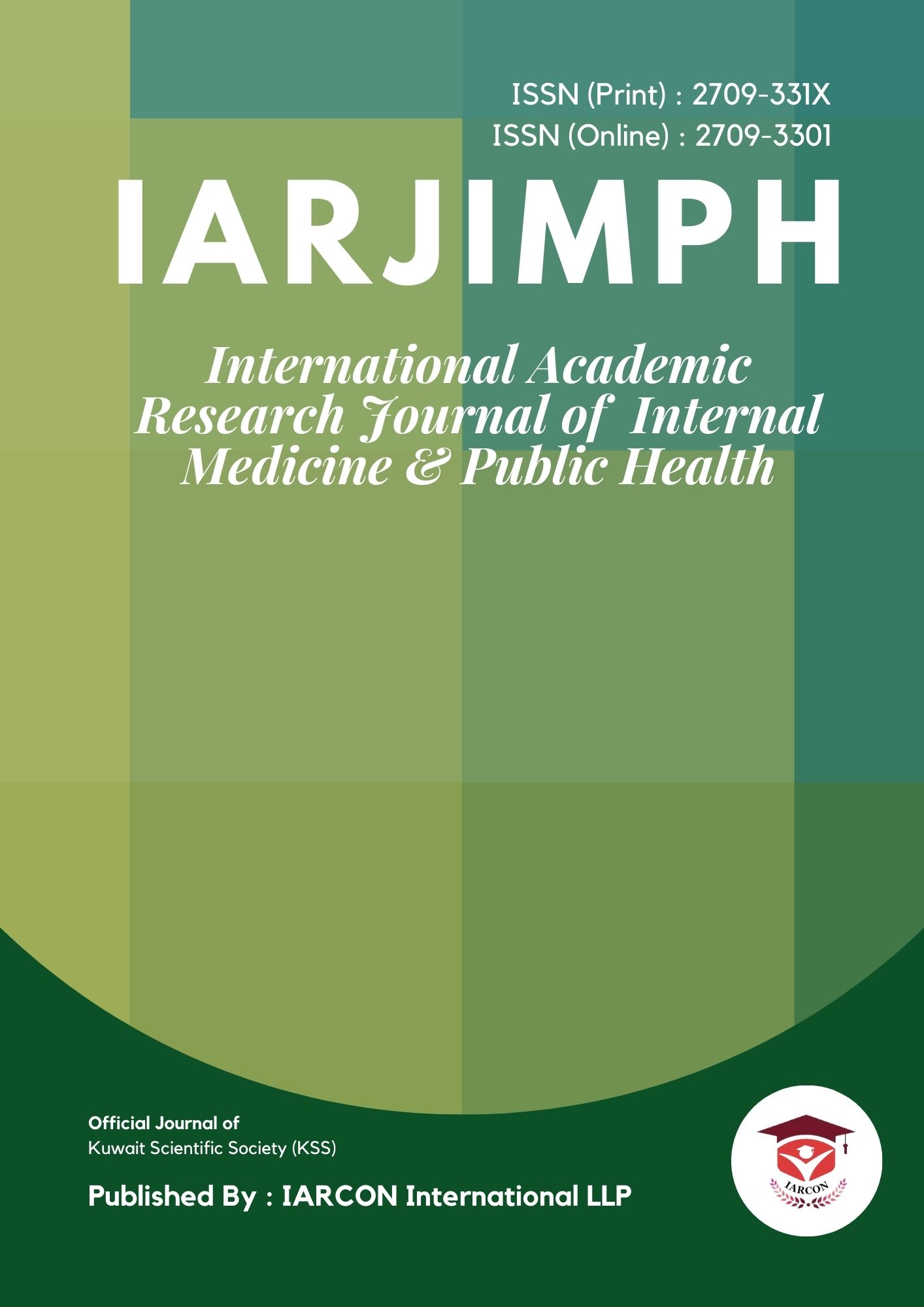
Niosome was introduced in 1978 by Lansone [1]. Niosome is a nonionic surfactants based on vesicle. They are formed mostly by nonionic surfactants and cholesterol incorporation as an excipient [2]. Niosome can be used as carrier of amphiphilic and lipophilic drug [3]. The niosome have ability to encapsulate different type of drug within their multi environmental structure. They present a structure similar to liposome and hence they represent alternative vesicular system with respect to liposome [4].The main aim of development of niosome is to control the release of drug in a sustained way, modification of distribution profile of drug and for targeting the drug to specific body site [1].There are different types of niosome multi lamellar vesicle (MLV), Large unilamellar vesicle (LUV) and Small unilamellar vesicle (SUV). There are many method of preparation of niosome such as Hand shaking, ether injection, bubbled method, ethanol injection and multiple membrane method. Generally the approach of niosome by various routes has reported. They are oral, parenteral, pulmonary, transdermal, vaginal and nasal routes. This review article represent the structure of niosome, advantages, disadvantages, method of preparation and characterization and factor affecting on formulation of niosome, zqapplication and types of niosome.
Niosomes is non-ionic surfactants based vesicle [2]. Niosomes is consider as one of the nanocarrier, which is considered because of their ability of drug loading efficiency is high and their biocompatibility. Niosomes is consider as non-ionic surfactants due to vesicular nano carrier madding which is used in targeted and sustained drug delivery system [5]. Niosomes was introduced in 1987 by 'Lancome'. In 1909, Paul ehrlich discovered new era in formulation of targeted drug delivery [1]. Niosomes is one of the potent drug nano particles [6]. In niosome non-ionic bilayer structures with hydrophilic core and a hydrophobic outer bilayer are embedded in a chitosan gel matrix. Chitosan is amino polysaccharide formed from alkaline deacylation of chitin polysaccharide [7]. Niosomes have been shown to keep many compound as measured by their entrapment efficiency [8]. During storage of niosomes has better instability during dispersion May be equivalent to that of liposome [9]. Depending upon the condition used the diameter of vesicle range from 50 to 1000 nm [3]. Principal of a targeted drug delivery system used possibility management in wide range of niosomes size. For example- of oral-less than 1000000 nm, at a local entered- less than 300 nm, at enter in a circulation blood - 10 nm [10]. Niosomes are stable nonionic microscopic vesicle consisting of one or several membrane of various structures. Wide application of nonionic surfactants and lipids in designing of such system is caused by their biocompatibility, ability and lower toxicity. [10] More than 50 different types of drug have been encapsulated in niosomes and administered inhalation, nasal, oral or parenteral route. Different method have been used for preparation of niosomes technique the ether injection method and the extensively used thin film hydration method [11].
Niosomes were first reported as a feature of cosmetic industry in the seventies and used in cosmetic formulation deviced by L'oreal. Niosome can be administered through various route such as oral, parenteral, topical, ocular, etc [12]. Chemically synthesized nonionic surfactants are amphiphilic compounds capable of reducing surface and interfacial tension and increase the solubility [15]. Nano niosomal drug delivery system after numerous advantage for this purpose. Nono niosome has an average particle size of 232 nm and had encapsulated 58% of the total protein present in the suspension [16].
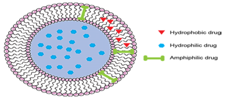
Figure 1: Schematic Representation of Niosomes
Structure
The niosome have hydrophilic head and hydrophobic tail sk the vesicle forming amphiphilic molecule. Polar heads facing hydrophilic region and non polar heads facing each other to form hydrophobic region (Figure 2) [3].
Composition and Components of Niosome
There are three major component used for the preparation of niosomes (Figure 3):
Cholesterol: Cholesterol is a steroid derivative usee to provide rigidity and proper shape and confirmation to niosome preparation [3]
Nonionic Surfactants: Span 60,40,20,80,tween 20,40,60,80 are nonionic surfactants used for the preparation oniosomes [4]
Cationic lipid with structures and physical properties clearly influence on the transfection efficiency and toxicity [13]
Features of Niosomes
There is no any special condition for storage and handling of Niosome
Niosome are the similar analogue to liposome [13]
In entrapped drug niosome have increase their stability and niosome are actively stable molecule or nano carrier. Niosome are reach site of action by oral, parenteral and topical route
Handling and storage of surface active agent to required any condition [13]
Niosome are osmatically active. It gives better availability to the particular site just by protecting the drug from biological environment [4]
Characteristics of Niosomes
Vesicle Diameter: Spherical structures of liposome and niosome are same, diameter of niosome can be determined by using light microscope and photon correlation optometry [13]
Entrapment Efficiency: Preparation of niosomal dispersion then entrapped drug is separated by using dialysis, centrifugation and gel chromatography [13]
Morphology: Niosome vesicle shape is spherical, laser light, scattering method is used to found mean diameter of niosome vesicle [9]
Membrane Rigidity: Measured by the availability of a fluorescence probe as a function of temperature [9]
Types of Niosomes
Following are the types of niosomes:
Niosome Made by Bola Surfactant: Niosome prepared by the surfactants and which are composed of omega- hexadecylbis-(1- aza -18 crown -6) (bola surfactants) [14]
Proniosome: Proniosome are the niosomal preparation excluding vesicle and surfactant [14]
Aspasome: Aspasome is a nanocarrier it formulated by addition of highly charge mixture like cholesterol palmitate and lipid diacetyl phosphate [14]
Vesicle in Water and Oil System (v/w/o): When aqueous niosome are emulsified in an oil phase shows in the formation of nanocarrier in water in oil emulsion. eg. Suspension formulated by the mixture of sorbitol monostirate, cholesterol and solulan C24 [13]
Other Types of Niosomes
Multilamellar Vesicle (MLV): Vesicle size is greater than 0.05 micro meter. This vesicle can be prepared by using Hand shaking method
Large Unilamellar Vesicle (LUV): Vesicle size is 0.025-0.05 micro meter. This vesicle are prepared by using sonication and extrusions method
Small Unilamellar Vesicle (SUV): Vesicle size greater than 0.10 micro meter. This vesicle are prepared by using reverse phase evaporation method
Applications of Niosomes
Niosome used as a drug carrier [14]
For control release of drug
To improve the stability and physical properties of the drug [14]
For targeting and retention of drug in blood circulation
Leishmaniasis- It is a plastic disease caused by protozoan parasite which used to invade the cells of the liver and spleen [14]
Used in transdermal drug delivery system [14]
Radio pharmaceuticals - It is the first application of niosome have been discovered by Erdogenet al in 1996 [15]
Used in parenteral application [7]
Advantages of Niosome
Niosome are used in different routes like oral,parenteral and topical in carriers of drug delivery [14]
In niosomes the handling and storage of surfactants required no any special condition [1]
They raised oral bioavailability of poorly absorbed drugs and enhance skin penetration of drugs [1]
Niosome increase the stability of entrapped drug [3]
They are osmatically active and stable [16]
They can be used in preparation for variety of drugs [3]
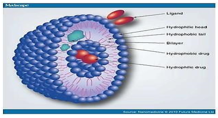
Figure 3: Composition and Components of Niosomes
Disadvantages of Niosome
Fusion [3]
Aggregation [3]
Physical instability [3]
Leaking of entrapped drug [3]
Need of intensive sonication lead to leakage of drug during storage [2]
Formulation and Evaluation of Niosome:
There are various method of preparation for niosome they are described as follows (Figure 4) [1]:
Multiple Membrane Extrusions Method: In this method the size of niosome can be reduced by passing them through membrane filter. This method can be used for production of multilamellar vesicle [1]
Ether Injection Method: This method provide a mean of making niosome by slowly introducing solution of surfactants dissolved in di ethyl ether into warm water maintain at 60°C.The surfactant mixture in ether in injected through 14 gauge needle into an aqueous solution of material [3]
Ultra-Sonication Method: Niosome were prepared by direct ultra-sonication of sample: 5mg of MB dissolved in 20 ml of hot deionized water at 80°C.The solution was added to a preheated surfactant and cholesterol. After hydrating the mixture for 60minutes. The hot distilled water was added to the system and make up final volume upto 100 ml. After 20minutes stirring at 500 rpm the system was sonicated using a probe sonicator. Then the prepared niosome were kept in fridge at 4-8°C for further characterization [17]
Ethanol Injection Method: This method is used for the preparation of small unilamellar vesicle (SUV) without sonication. In this method, ethanol solution of surfactants is injected rapidly through a fine needle into excess of saline and aqueous medium. Vaporization of ethanol leads to the formation of vesicle [4]
Bubbled Method: The bubbling unit consists of round bottom flask with three necks positioned in water bath to control the temperature. Water cooled reflux and temperature is positioned in the first and second neck and nitrogen is supplied through the third neck. Cholesterol and surfactant together in buffer pH 7.4 at 70°C, the dispersion is mixed for 15second, with high share homogenizer and immediately afterwards bubbled at 70°C using nitrogen gas [2]
Evaluation of Niosome
Entrapment Efficiency: After preparation of niosomal dispersion, unentrapped drug is separated by dialysis, centrifugation and gel filtration method [18]
Vesicle Diameter: Niosome are similar to liposome, niosome have spherical shape so their diameter can be determined by using light microscopy [18]
Invitro Release: In this method rate study include the use of dialysis tubing. The vesicle suspension is peppited into a bag made up of tubing and sealed. The bag containing the vesicle is placed in 200ml of buffer solution in a 250ml beaker with constant shaking at 25°C [18]
Factors Affecting on Formulation of Niosome
Drug: Entrapment of drug in niosomes increase vesicle size, probably by interaction of solute with surfactant head group, to increase change of surfactants [2]
Hydration Temperature: The size and shape of the niosome are affected by the temperature of hydration [5]
Cholesterol Contents: Incorporation of cholesterol increase the entrapment efficiency and hydro dynamic diameter of niosome [5]
Amount and Type of Surfactants: The mean size of niosome increase proportionally with increase in the HLB surfactant like span 85 (HLB 1.8) to span 20 (HLB 8.6) because the surfactants free energy decrease with an increase in hydrophobicity of surfactants [2]
Charge: Presence of charge leads to an increase in interlamellar distance between successive bilayers in multi lamellar vesicle structures [5] (Figure 5)
Figure 4: Formulation of Niosomes
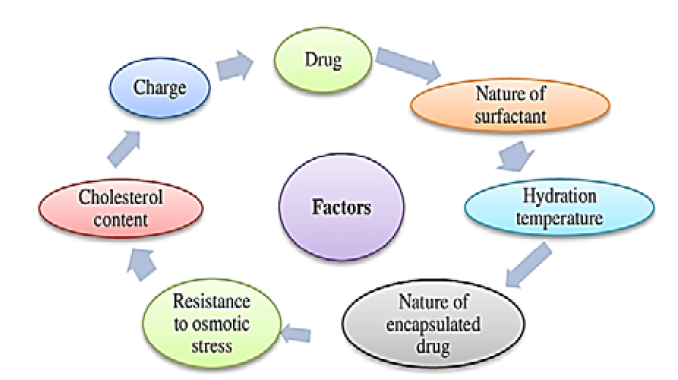
Figure 5: Factors Influencing on Niosomal Formulation
Surfactant Used in Niosomes
Ether Linked Surfactant: In this surfactant hydrophilic and hydrophobic moiety are ether linked. Polyoxyethelyene alkyl ether (CnEOm) where n is number of carbon atoms varies between 12 and 18 and m is the number of oxyethylene unit varies between 3 and 7 [18] (Table 1)
Di Alkyl Chain Surfactant: Surfactant was used as a principal component of niosomal preparation of stibogluconate [18]
C16H33CH-O[-CH2-CH-O]7-H
|
CH2 CH2OH
|
C12H25-O (mol. Wt. 972)
Ester Linked Surfactant: These are the surfactants in which hydrophilic and hydrophobic moieties are ester linked. Ester linked surfactant
C15H31CO[O-CH2-CH-CH2]2-OH
|
OH (mol. Wt. 393)
This surfactant used in delivery of sodium stibogluconate to the experimental marine visceral leishmaniasis following administration of niosomal system. The commercial sorbitan esters are H-C-OH mixtures of the partial easter of sorbitol [18]
Sorbitol Esters
CH2
|
H-C-OH
|
RCOO- C-H
|
H-C-OH
|
H-C-OOC-R
|
CH2OOC-R
Where R is H or an alkyl chain.
The formula of representative component is shown above. Sorbitan esters based niosome bearing methotrexate were prepared and evaluated for pharmacokinetic of the entrapped methotrexate in tumor bearing mice [18]
Poly Sorbates: The typical structural formula for polysorbates is [18]
CH2
|
H-C-O(CH2-CH2-O) H
|
(OCH-CH2)-O-C-H
|
H-C-O-(CH2-CH2-O)y H
|
CH2-O(CH2-CH2-O)z OCR
After extracting by soxhlet, the resulting extract was loaded in the nano niosome system by thin film method and was subjected to physical, chemical and cellular transporter [8].
Niosome Composition with Liposome
Single chain unchanged surfactant and cholesterol are usee for the preparation of niosome and double chain phospholipid that are neutral or charged are used for the preparation of liposome. Niosome behave like liposome inside our body [14]. Niosome and liposome are functionally same, they having same physical properties and act as amphiphilic vesicle. Both are used in targeted and sustained drug delivery system. Niosome shows better chemical stability as compared to liposome [19].
Stability and Toxicity of Niosome
Niosome surfactant which is used in niosomes nontoxic and have been no toxic effect on animal studies during use of niosome as drug carrier. Niosome are stable structure so it is known as stabilized niosome also [13]. The only reason that May lead to instability of niosome is the nature of surfactants used in its formation but till now no such report available on the in vivo toxicity of niosome linked with the concentration of ether or ester surfactant used in the preparation of vesicle [20].
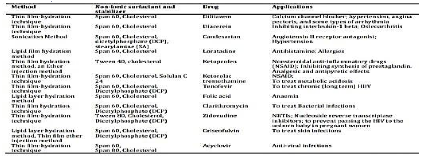
Table 1: Methods of Preparation with Various Non-Ionic Surfactants and Stabilizer with its Applications
Factors Affecting on Stability and Toxicity of Niosome
Which type of surface active agent we used.
Storage
Temperature
Detergent
Encapsulated drug nature [15]
Routes of Administration
Nano niosome are currently user as vesicle drug delivery system with many route of administration such as oral route, ophthalmic, transdermal, parenteral, pulmonary, vaginal and nasal routes:
Oral Route
The oral delivery of recombinant human insulin using niosomal formulations was demonstrated by a study involving polyoxyethylene alkyl ethers based niosomes. Significantly higher protection activity was seen in Brij 92/cholesterol (7:3 molar ratios) in which only 26.3±3.98% of entrapped insulin was released during 24 h in simulated intestinal fluid (SIF) 10. Niosomes were prepared from Span40, Span60, and Cholesterol using reverse evaporation method. Niosomal formulation could be promising delivery system for gliclazide with improved bioavailability and prolonged drug release profile. Gliclazide-loaded niosomes were formulated and evaluated for their in-vitro as well as in-vivo characteristics in an attempt to improve the oral bioavailability. The in-vitro release studies of drug from niosomes exhibited a prolonged drug release as observed over a period of 24 hrs24 [1].
Parenteral Route
On the basis of these pharmaceutical and clinical facts, innovative niosomes made up of α,ω-hexadecyl-bis-(1-aza-18-crown-6) (bola) [9] (Figure 6).
Ophthalmic Route
To minimize the problem associated with conventional eye drops, different ocular drug delivery devices have been used [9].
Pulmonary Route
Nanocarriers could be used for protection and more effective delivery of different therapeutics to respiratory tract.Nanocarriers could be used for protection and more effective delivery of different therapeutics to respiratory tract [9].
Nasal Route
Priperm et al. Prepared melatonin encapsulated niosomes composed of Span 60/CHOL/sodium deoxycholate [9].
Transdermal Route
Preparation of vesicles could be a versatile technique to enhance topical penetration of applied drugs trough natural barrier layer, stratum corneum. Alvi et al. (65) reported that vesiculization of 5-FU not only improved the topical delivery, but also enhanced the cytotoxic effect of 5-FU in actinic keratosis and non-melanoma skin carcinoma. (66) used Span 60, Span 20, and Tween 20 with CHOL to prepare nano size vesicle of minoxidil. Niosome formulation prepared with 1:2 ratio of Span 60 and CHOL showed 17.21±3.2 % skin retention of minoxidil, which was six fold more than that of minoxidil gel as control. By preparation of nano-niosomes, both poor water solubility and low skin penetration of minoxidil were addressed.
In addition to nanovesicle encapsulation, some other methods were developed for enhancing transdermal transport of large molecules such as insulin [9]
Vaginal Route
They concluded that vaginally administrated nano-niosomes might be a good carrier for protein drugs such as insulin.
Two types of insulin vesicles with Span 40 and Span 60 were prepared by lipid phase evaporation and sonication methods with particle sizes of 242.5 nm and 259nm.
Approach of Niosome
The approach of niosome by various routes has been reported. They are oral route delivery, transdermal delivery, delivery of peptide drug, harm one delivery, cosmetic delivery:
Oral Route Delivery: The effective delivery of drug the oral delivery system is used. Niosome are prepared from span 40,span 60 and cholesterol using reverse evaporation method [1]
Transdermal Delivery: Slow preparation is a disadvantage of transdermal route of delivery. When we increase the penetration rate the transdermal delivery of drug incorporated in niosomes [1]
Cosmetic Delivery: Niosome used in cosmetic and skin care application including their ability to enhance the stability of entrapped drugs , better bioavailability of poorly absorbed piece [1]
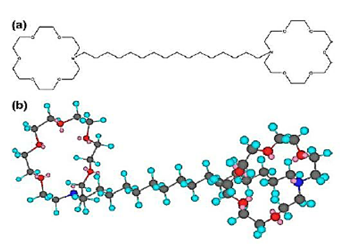
Figure 6: Chemical Structure of Bolaform Surfactants: (a)
Bola A-16, (b) Bola C-16 (54)
Formation of Niosome
Niosome are formed such as sorbitol is coated with surfactant. The dry formulation of water soluble carrier particles that are coated with surfactant. This process of preparation is known as Proniosome. The niosome are recognized by the addition of aqueous phase T>™ and brief agitations. Formulation of niosome from maltodextrine which is used a production of Proniosome [21].
Characterization and Aspects Affecting Formulation of Niosome
Nature of Surfactant: Niosome can be prepared by lipophilic tail and lipophilic head. In the alkyl chain length of surfactant with the range of C12-C18 are most appropriate for formation of noisome [14]
Composition of Membrane: The bilayered surface of niosome were directly effect on the charge and rigidity properties like physical and chemical property of the drug present in the encapsulated form [14] (Table 2)
Niosome Purification and Separation of Unentrapped Drug
The purification of niosome suspension was carried out by using two different method - dialysis, gel permeation method and centrifugation method.
Dialysis: Loaded niosome suspension were placed in dialysis bag and let floating in the corresponding hydration media in a 1-100v/v ratio. Dialysis time was adjusted depending upon the encapsulated compound. Reverse phase HPLC was used to determined the concentration of free compound in the collected sample [21]
Gel Permeation Method: AA and RB loaded niosome were purified using a sephadex G-25 superfine column while B12 and RB loaded niosome in PEG, were purified using a gravity elution PD column packed with sepharose CL4B. Sepharose CL4B was used since in optimization step.Control solution of the corresponding encapsulated compound in the selected media were used to assess the efficiency of the applied method [21] (Table 3)
Table 2: Characterization Parameters of Niosome

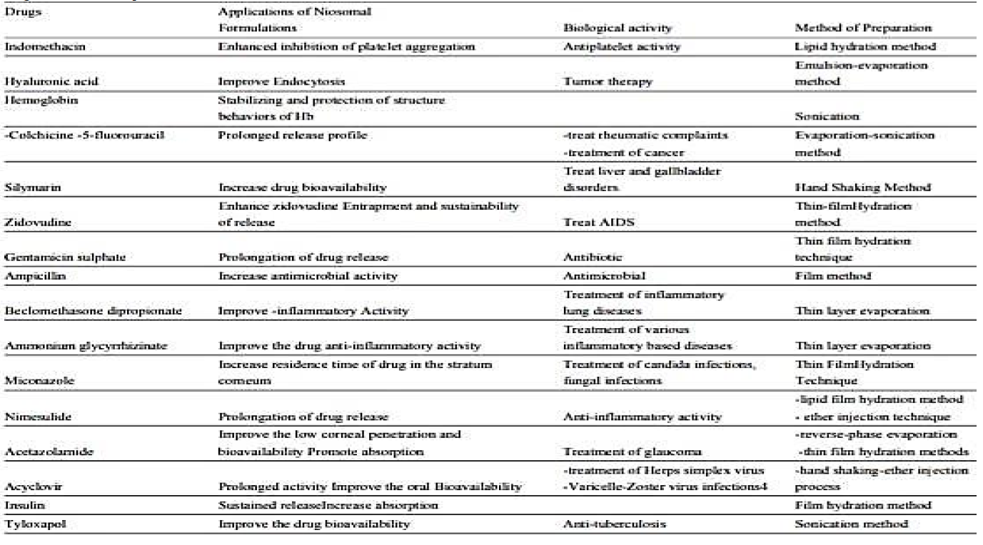
Table 3: Drugs Used For Preparation of Niosome
Niosome are more stable than liposome and immense ability to alter pharmaceutical profile of antileishmanial and anti-cancer drugs. Niosome have been proven to useful control drug delivery system for different routes such as transdermal, oral, parenteral and ophthalmic routes. Niosome represent a promising drug delivery module. They present a structure similar to liposome and hence they can represent alternative vesicular system with respect to liposome due to the niosome ability respect to encapsulated different type of drugs. Niosomal drug delivery system is one of the best example of great evolution in drug delivery technologies and nano technology. Niosome have very important and key role in various types of drug delivery like targeting, topical, ophthalmic and parenteral. Niosome are very useful in bright future for pharma industries. The function of niosome as targeted drug delivery system has been studied and new classes of niosome have been developed. Niosome characteristics particularly entrapment efficiency may be affected by preparation technique along with the nature of the incorporated drugs, cholesterol content and the type of wetting agent. Niosome are brodly used for topical drug delivery enhancing the permeation and sustaining the release of drugs.
Ali, Nasir and Harikumar, S.L. “Niosome: An excellent tool for drug delivery.” International Journal of Research in Pharmacy and Chemistry, vol. 2, no. 2, 2012, pp. 2231–2781.
Gurjar, Pravin et al. “Niosome: A promising pharmaceutical drug delivery.” International Journal of Pharmaceutics and Drug Analysis, vol. 2, no. 5, 2014, pp. 425–431.
Sharma, D., Ali, A.A.E. et al. “Niosome as novel drug delivery system: review article.” Pharma Tutorial, vol. 6, no. 3, 2018, pp. 58–65.
Gurjar, Pravin and Nike, Nivedita. “Niosome: A promising pharmaceutical drug delivery.” International Journal of Pharmacy, vol. 2, no. 5, 2014, pp. 425–431.
Kour, D. and Kumar, S. “Niosome: Present scenario and future aspects.” Journal of Drug Delivery and Therapeutics, vol. 8, no. 5, 2018, pp. 35–43.
Moghassemi et al. “Growth factor loaded nano niosome gel formulation and characterization.” AAPS PharmSciTech, vol. 17, no. 10, 2016, p. 1280.
Wiranowska, M. et al. “Preferential drug delivery to tumor cells than normal cells using a tunable niosome chitosan double package nano delivery system: A novel in vitro model.” Cancer Nanotechnology, vol. 11, no. 3, 2020.
Bartelds, R. et al. “Niosome: An alternative for liposomal delivery.” PLOS One, vol. 13, no. 10, 2018, p. 1371.
Mothilal et al. “Niosome as an emerging formulation tool for drug delivery: A review.” International Journal of Applied Pharmaceutics, vol. 11, no. 2, 2019, pp. 7–15.
Diskaeva, E.I. and Vecher, O.V. et al. “Particle size analysis of niosome as a function of temperature.” Nanosystems: Physics, Chemistry, Mathematics, vol. 9, no. 2, 2018, pp. 290–294.
Khan, D.H. et al. “Formulation optimization and in vitro characterization of rifampicin and ceftriaxone dual drug loaded niosome with high energy probe sonication technique.” Journal of Drug Delivery Science and Technology, vol. 58, 2020, p. 101763.
Yaakob, Harisun and Bagheri, Ali. “Niosomal drug delivery system: Formulation, preparation and applications.” World Applied Sciences Journal, vol. 32, no. 8, 2014, pp. 1671–1685.
Patino, B. et al. “A novel niosome encapsulated essential oil formulation to prevent Aspergillus flavus growth and aflatoxin contamination of maize grains during storage.” Toxins, vol. 11, 2019, p. 646.
Jain, Siddharth C. et al. “A brief review on niosomes.” Journal of Pharmacy Research, vol. 11, no. 5, 2017, pp. 450–458.
Ullah, S., Razashah, M. et al. “Development of a biocompatible creatinine-based niosomal delivery system to enhance oral bioavailability of clarithromycin.” Drug Delivery, vol. 23, no. 9, 2016, pp. 3480–3491.
Moghassemi et al. “Growth factor-loaded nano niosomal gel formulation and characterization.” AAPS PharmSciTech, 2016.
Ali, F. and Akbari, J. et al. “Methylene blue-loaded niosome: preparation, physicochemical characterization and in vivo wound healing assessment.” Drug Delivery and Translational Research, vol. 10, 2020, pp. 1428–1441.
Tangri, Pranshu and Khurana, Shaffi. “Niosome: formulation and evaluation.” International Journal of Biopharmaceutics, vol. 2, no. 1, 2011, pp. 47–53.
Hamishekhar, H. et al. “Niosome as a propitious carrier for topical drug delivery.” Expert Opinion on Drug Delivery, vol. 10, no. 2, 2013, pp. 261–272.
Nazis, Khanam et al. “Recent trend in drug delivery by niosome: A review.” Asian Journal of Pharmaceutical Research and Development, vol. 1, no. 3, 2013, pp. 115–122.
Singh, Soumya. “Niosome: A role in targeted drug delivery system.” International Journal of Pharmaceutical Science and Research, vol. 4, no. 2, 2013, pp. 550–557.
