Herpes Zoster Ophthalmicus is caused by reactivation of varicella zoster virus.It involves eye causing conjunctivitis, keratitis, uveitis, keratitis and rarely cranial nerve palsy and optic neuritis. Here we report case of Herpes Zoster keratouveitis with oculomotor nerve palsy in diabetic patient.
Keywords
Herpes Zoster Ophthalmicus
Oculomotor Nerve Palsy
Uveitis
INTRODUCTION
Varicella zoster virus (VZV) is a herpes virus which involves mainly the younger age group showing a higher infectivity rate.
It manifests typically in two distinct forms: varicella (chickenpox) and herpes zoster (shingles). Primary VZV presents as diffuse vesicular rash whereas the reactivation of infection in previously infected individuals causes herpes zoster (HZ), which presents as localized, painful dermatomal rash [1].
Main risk factors for its reactivation are advanced age and suppressed immune state. Spinal cord dorsal ganglia act as the main reservoir .It mainly occurs in temperate regions with highest cases presenting during the summer season. Herpes zoster ophthalmicus (HZO) mainly spreads from theGasser ganglion involving the V1 territory of trigeminal nerve. It mainly involves around 10%–20% of all the herpes zoster cases [2].
Herpes zoster ophthalmicus (HZO) can cause maculo-vesicular rash , keratitis, uveitis, retinitis, and rarely cranial nerve palsies and optic neuritis. We report a case of herpes zoster ophthalmicus with oculomotor palsy involving the pupi [1].
Case Report
A 67-year-old diabetic patient was referred from the medicine ward for ophthalmology opinion. He presented with complains of painful lesions around the righteye and was not able to open the eye for the past 7 days.
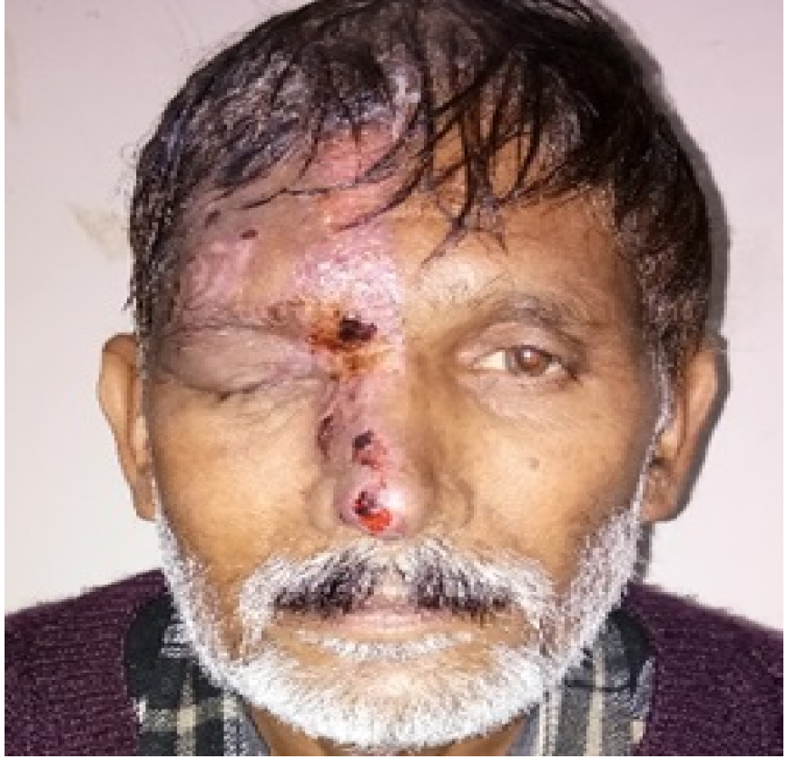
On local examination, a vesicular rash with crusting was seenon the left side of forehead involving the right upper eyelid and tip of the nose; respecting vertical meridian (Figure 1).
Figure 1: Showing Vesicular Rash Involving Right Forehead, Ptotic Lid and Nose Involving Nose Tip
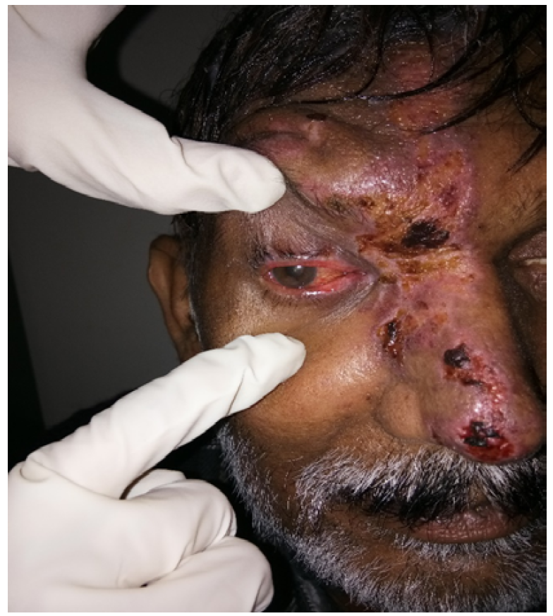
On further ophthalmic examination, itshowed severeptosis, and eye ball was out and down; visual acuity wasfinger counting at 1 foot in right eye and 6/9 in left eye. Pupil wasfixed dilated in right eye and normal in left eye. The extraocular movementswere restricted in elevation and adduction in right eye and full in left eye (Figure 2).
Figure 2: Showing Down and Out Eye Ball, Keratitis and Conjunctivitis with Fixed Mid Dilated Pupil
On slit lamp examination, conjunctiva wascongested, cornea had stromal infiltration involving lower cornea, KPs with 3+ anterior chamber reaction and 1+ vitritis.Retina was within normal limits. Intraocular pressure was normal. Patient was having right eye keratouveitis (herpes zoster ophthalmicus) with third nerve palsy with pupillary involvement.
Patient was started on tablet valcyclovir 1gm TDS, eye drop moxifloxacin and dexamethasone, eye drop cyclopentolate and was reviewed after one week.On follow up examination, stromal infiltration decreased with no anterior chamber reaction; but no improvement of ptosis was seen. After 1 month follow up, cornea shows minimal scarring with improvement in ptosis and extraocular movements.
DISCUSSION
VZV reactivation usually involves the thoracic dermatome and 5th cranial nerve. Incidence of 5th cranial nerve in herpes zoster ophthalmicus is around 5-29%.
A cranial nerve III palsy results in adduction and vertical ocular motility deficits and it may involve the pupil [3,4]. Partial palsies can result in varying degrees of involvement of directions of eye movements. Causes are varied and can include space-occupying lesions, microvascular infarctions, aneurysmal compressions, inflammation, infections, and trauma.
Typically, in HZO, the virus causes a dermatitis/vesicular rash of the ophthalmic branch of CN V. Corneal involvement is a relatively common ocular finding and can lead to pseudo-dendritic keratitis which can result in decreased corneal sensitivity, neurotrophic ulcers, and/or decreased vision secondary to scarring [5]. If the tip of the nose is involved with the vesicular rash this is termed “Hutchinson's sign” and suggests there is an increased risk of corneal/ocular sequelae secondary to nasociliary nerve involvement [6]. One of the most common complications of any HZ outbreak tends to be post-herpetic neuralgia (PHN) which can cause very intense episodes of pain affecting the same areas as the vesicular rash. But cranial nerve palsies are rare.
CONCLUSION
Herpes zoster Ophthalmicus mainly involve the skin and the cornea, cranial palsies are rare complications, which need to be detected early and treated effectively so as to avoid long term defects.
REFERENCE
Pascual-Prieto, J., et al. “Parálisis oculomotoras en el contexto de una reactivación del virus varicela-zóster: a propósito de 4 casos.” Archivos de la Sociedad Española de Oftalmología year 2020.
Albrecht, M.A. “Clinical features of varicella-zoster virus infection: chickenpox.” UpToDate, edited by Connor, R.F., Wolters Kluwer, accessed May 23, 2024.
Savino, P.J., and Danesh-Meyer, H. “Color atlas and synopsis of clinical ophthalmology (Wills Eye Hospital): neuro-ophthalmology.” McGraw-Hill, 2003, pp. 152–160, 202–209, 236–240.
Penne, R.B. “Color atlas and synopsis of clinical ophthalmology (Wills Eye Hospital): oculoplastics.” McGraw-Hill, 2003, pp. 80–81, 82–83, 86–87, 152–157, 249–251.
Sampathkumar, P., et al. “Herpes zoster (shingles) and postherpetic neuralgia.” Mayo Clinic Proceedings vol. 84, year 2009, pp. 274–280.
Harthan, J.S., and Borgman, C.J. “Herpes zoster ophthalmicus-induced oculomotor nerve palsy.” Journal of Optometry vol. 6, no. 1, 2013, pp. 60–65. doi: https://doi.org/10.1016/j.optom.2012.08.005
Recommended Articles
Research Article
Knowledge of the Patient's Family about the Level of Emergency Based on Triage with the Satisfaction of the Patient's Family in the Emergency Department of the Sumedang Area General Hospital
Roganda Situmorang,
...
Raida Rihadatul Aisyi
Published: 30/03/2024
Download PDF
Research Article
The Influence of Murottal Al Qur'an Surat Ar Rahman Irama Hijaz Therapy on the Pain Scale of Post-Explorative Laparatomy Patients in the Icu Room of Bandung City Hospital
Andri Nurmansyah,
...
Fifi Siti Fauziah Yani
Published: 30/03/2024
Download PDF
Research Article
The role of IFN-g gene polymorphism (+874 T/A) as predisposing factor for pulmonary tuberculosis
Download PDF
Research Article
Assessment of Knowledge on Common Pediatric Infections and Their Prevention Among Parents in Chamba, Himachal Pradesh
Isha Bhatia,
Rupika
Published: 20/12/2024
Download PDF
PO Box 101, Nakuru
Kenya.
Email: office@iarconsortium.org
Editorial Office:
J.L Bhavan, Near Radison Blu Hotel,
Jalukbari, Guwahati-India
Katoch, K., Sharma, P., Sharma, A. & Poonia, K. (2024). Oculomotor Nerve Palsy in Herpes Zoster Ophthalmicus. Scientific Research Journal of Clinical and Medical Sciences, 4(2), 1-2.
MLA
Katoch, Kritika, et al. "Oculomotor Nerve Palsy in Herpes Zoster Ophthalmicus." Scientific Research Journal of Clinical and Medical Sciences 4.2 (2024): 1-2.
Chicago
Katoch, Kritika, Pankaj Sharma, Aakanksha Sharma and Kavita Poonia. "Oculomotor Nerve Palsy in Herpes Zoster Ophthalmicus." Scientific Research Journal of Clinical and Medical Sciences 4, no. 2 (2024): 1-2.
Harvard
Katoch, K., Sharma, P., Sharma, A. and Poonia, K. (2024) 'Oculomotor Nerve Palsy in Herpes Zoster Ophthalmicus' Scientific Research Journal of Clinical and Medical Sciences 4(2), pp. 1-2.
Vancouver
Katoch K, Sharma P, Sharma A, Poonia K. Oculomotor Nerve Palsy in Herpes Zoster Ophthalmicus. Scientific Research Journal of Clinical and Medical Sciences. 2024 Jul;4(2):1-2.



 Download PDF
 Download XML