Contents
Download PDF
pdf Download XML
182 Views
9 Downloads
Share this article
Research Article | Volume 4 Issue 2 (July- Dec, 2024) | Pages 1 - 3
DIGITAL PANORAMIC RADIOGRAPHIC STUDY OF MENTAL FORAMEN (LOCATION AND TYPE) IN HIMACHAL PRADESH POPULATION
 ,
 ,
1
MDS, Medical Officer Dental, Dept of Health and Family Welfare, H.P, India
2
Postgraduate student and Research Scholar, Department of Oral Medicine and Radiology, HPGDC, Shimla, India
Under a Creative Commons license
Open Access
Received
June 5, 2024
Revised
July 20, 2024
Accepted
July 29, 2024
Published
Aug. 16, 2024
Abstract

Introduction: Assessment of MF specially position is critical for successful anaesthetic and surgical procedures. AIM: Present study was to assess the most common type and position of MF in Himachal Pradesh population. Material And Methods: Retrospective study was done using 326 digital Orthopantomogram (OPG), patients age ranging from 16 to 65 years. Location and type of MF are identified by tracing inferior alveolar canal and position and type is classified accordingly. Results: The most common location of MF on the both sides were found to be Location “2” i.e. below second Premolar accounting to be 55.50%. No MF was seen in position 3 and 4 i.e. below the Canine and below the first Molar were evident. The most common type of MF seen was type 2 i.e. Separate type, found in 157 subjects accounting for 48.16% on both sides. No subject radiograph with type 4 MF i.e. Unidentified type was seen. Conclusion: The study concluded that there was significant difference in the location and type of mental foramen among Himachal Pradesh population.

Keywords
INTRODUCTION

The Mental Foramen (MF) is an opening found in the body of mandible which represents the termination of Inferior Alveolar Canal which opens and Mental nerve passes through MF and supplies sensory innervations and nutrition to the chin, lower lip and gingiva, teeth on the ipsilateral side of the mandible1. The foramen is hard to be directly visualized or palpated hence radiographic evaluation of the position of MF becomes a mandatory for accurate diagnosis. The exact position assessment of MF is critical for both clinical and diagnostic procedures. The nerves and vessels could be injured during mandibular fixation/reduction, periapical surgery, orthognathic surgery or implant placement. Permanent injury may cause paraesthesia or anaesthesia on the affected side for the rest of his/her life2.
 

On radiographs, MF appears as a radiolucent area mostly having a circular or oval shape. Its visualization on intraoral radiograph (IOPA) can be difficult, due to its position below the edge of the film. Patient with the large mandibular tori and a shallow floor of mouth or crowded teeth may prevent the proper placement of film during radiographic examination3. Hence, the best radiographs that can show MF are Orthopantomogram (OPG), cone-beam computed tomography (CT), and CT scan4. OPG being most convenient and economic of all.


As the MF has been reported to differ in position and type in different population, this study was aimed to evaluate these variables among Himachali patients.

MATERIAL AND METHODS

A Retrospective study on total 326 digital Orthopantomogram (OPG), patients age ranging from 16 to 65 years was done. These OPGs were taken of patients visiting for other dental check-ups and procedures.

Inclusion Criteria: 

  1. Any patients aged above 18 years subjected to OPG for various purposes such as orthodontic assessment, Wisdom tooth assessment, dentition assessment, and prosthetic assessment etc. 

  2. Good quality image (optimal angulation etc).

  3. Dentate patients, especially with erupted mandibular canines, premolars and first molars.


Exclusion criteria 

  1. Patients with hereditary facial asymmetries, congenital anomaly in the mandible that could affect the interpretation of radiographic image.

  2. History of surgical interventions on the lower jaw.

  3. Presence of pathologies or fracture line in the lower jaw.

  4. History of orthodontic treatment.

  5. Radiographs of low quality.

  6. Patients under the age of 18.


The mental foramen was located as radiolucency, traced by following the inferior alveolar canal.


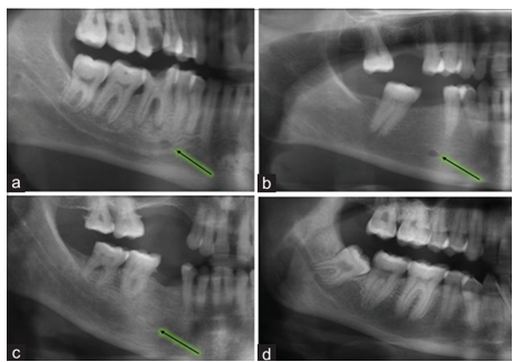
Appearance of Mental foramen (MF) can be classified in four types5:

  1. Type 1: Continuous type: in continuity with the mandibular canal.

  2. Type 2: Separated type: separated from mandibular canal.

  3. Type 3: Diffuse type: indistinct or ill-defined border.

  4. Type 4: Unidentified type: the foramen which could not be seen on radiograph.


Appearance of mental foramen on OPG (a) Type I – continuous type (b) Type II – separate type (c) Type III – diffuse type (d) Type IV – unidentified type5


The Location of the mental foramen on radiograph can be classified as:

  1. Situated below the first premolar. 

  2. Situated below the second premolar. 

  3. Situated below the Canine.

  4. Situated below the first Molar.

  5. Situated between Premolars. 

  6. Situated between second Premolar and first Molar.

  7. Situated between first Premolar and Canine.

RESULTS

Total 326 OPGs were examined which fulfilled the inclusion criteria, out of which 200 belonged to males and 126 to females (table 1).


S. NO

SUBJECTS

NUMBER

PERCENTAGE

1

Male

200

61.35%

2

female

126

38.65%

Table 1


Age: subjects ranging from 18 to 65 were selected. Mean age of males was 37.77 ± 1.03 years and of females was 34.77 ±1.22 years, difference was statistically insignificant (table2).


S. NO

SUBJECTS

AGE±SD

1

Male

37.77 ± 1.03 Y

2

female

34.77 ±1.22 Y

Table 2


Position/ Location of Mental Foramen: The most common location of MF on the both sides were found to be Location “2” i.e. below second Premolar accounting to be 55.50%. second most common position was “5” i.e. between both Premolars, with frequency of 27% on both sides. Third most common was position “9” with frequency of 8.30% on both sides. Frequency of occurrence in location “1” i.e. below first premolar and location “6” i.e. below second premolar and first Molar were mere 6.10% and 8.30% on both sides. No MF was seen in position 3 and 4 i.e. below the Canine and below the first Molar were evident (table 3).


S.No

POSITION

TOTAL NUMBER

PERCENTAGE

  1.  

Situated below the first premolar. 

20

6.10%

  1.  

Situated below the second premolar. 

181

55.50%

  1.  

Situated below the Canine.

0

0%

  1.  

Situated below the first Molar.

0

0%

  1.  

Situated between Premolars. 

88

27.00%

  1.  

Situated between second Premolar and first Molar.

10

3.06%

  1.  

Situated between first Premolar and Canine.

27

8.30%

Table 3


Type of MF: the most common type of MF seen was type 2 i.e. Separate type, found in 157 subjects accounting for 48.16% on both sides. Second most common is type 1 i.e. Continuous type with number 92 and frequency of 28.22%. Third most common is type 4 i.e. Diffuse type with number 77 and frequency of 23.62%. No subject radiograph with type 4 MF i.e. Unidentified type was seen (table 4).


S.No

POSITION

TOTAL NUMBER

PERCENTAGE

  1.  

Situated below the first premolar. 

92

28.22%

  1.  

Situated below the second premolar. 

157

48.16%

  1.  

Situated below the Canine.

77

23.62%

  1.  

Situated below the first Molar.

0

0%

Table 4

DISCUSSION

Mental foramen cannot be assessed externally by palpation or otherwise, making it mandatory to assess MF through radiographic methods6. Easiest and most convenient is the orthopantomogram (OPG). Identification of MF is of vital importance as many procedures surgical and anaesthetic require this information for their flawless and successful execution7.


In our study we found that the most common location of MF on the both sides were found to be Location “2” i.e. below second Premolar accounting to be 55.50%. second most common position was “5” i.e. between both Premolars, with frequency of 27% on both sides. Third most common was position “9” with frequency of 8.30% on both sides. Frequency of occurrence in location “1” i.e. below first premolar and location “6” i.e. below second premolar and first Molar were mere 6.10% and 8.30% on both sides. No MF was seen in position 3 and 4 i.e. below the Canine and below the first Molar were evident. Also, the most common type of MF seen was type 2 i.e. Separate type, found in 157 subjects accounting for 48.16% on both sides. Second most common is type 1 i.e. Continuous type with number 92 and frequency of 28.22%. Third most common is type 4 i.e. Diffuse type with number 77 and frequency of 23.62%. No subject radiograph with type 4 MF i.e. Unidentified type was seen.


It was also observed that both sides i.e. right and left side of the mandible showed same location and type of MF, no discrepancy was noticed.

CONCLUSION

In our study we found that most common location of MF was observed to be below second Premolar and most common type of MF was separate type.


These findings can be used as reference by the Dentists, while performing clinical procedures involving mental foramen on Himachali population.


Conflict of Interest: None.

Financial disclosure: None.

REFERENCES
  1. Gupta, V., Pitti, P., and Sholapurkar, A. "Panoramic Radiographic Study of Mental Foramen in Selected Dravidians of South Indian Population: A Hospital Based Study." Journal of Clinical & Experimental Dentistry, vol. 7, no. 4, 2015, pp. e451-6. doi:10.4317/jced.52373.

  2. Phillips, J. L., Weller, R. N., and Kulild, J. C. "The Mental Foramen: I. Size, Orientation, and Positional Relationship to Mandibular Second Premolar." Journal of Endodontics, vol. 16, 1990, pp. 221-3. doi:10.1016/S0099-2399(06)80434-7.

  3. Afkhami, F., Haraji, A., and Boostani, H. R. "Radiographic Localization of the Mental Foramen and Mandibular Canal." Journal of Dentistry (Tehran), vol. 10, 2013, pp. 436-42. doi:10.22038/jdmt.2013.2815.

  4. Elhassan, A. T., Musrati, A. A., Ehtiwash, N. M., Peeran, S. W., and Alzahrani, F. M. "Location of Mental Foramen in a Group of Libyan Patients: An Orthopantomographic Study." Dental Medicine Research, vol. 7, 2019, pp. 45-8. doi:10.4103/dmr.dmr_13_19.

  5. Asrani, V. K., and Shah, J. S. "Mental Foramen: A Predictor of Age and Gender and Guide for Various Procedures." Journal of Forensic Sciences & Medicine, vol. 4, 2018, pp. 76-84. doi:10.4103/jfsm.jfsm_10_18.

  6. Ali, A., Shah, A., Hakim, T., Shawl, J., Bhat, I., and Aslam, S. "Panoramic Radiographic Study of Mental Foramen in Selected Kashmiri Population." Annals of International Medical and Dental Research, vol. 2, no. 4, 2016, pp. 112-14. doi:10.21276/aimdr.2016.2.4.24.

  7. Dua, N., Sharma, P., Kaur, H., Kaur, M., and Girdhar, M. "Radiographic Study of the Appearance, Location, and Size of Mental Foramen on Digital Panoramic Radiographs." Archives of Dental Research, vol. 12, no. 2, 2022, pp. 109-14. doi:10.5334/adr.2587.

Recommended Articles
Research Article
Knowledge of the Patient's Family about the Level of Emergency Based on Triage with the Satisfaction of the Patient's Family in the Emergency Department of the Sumedang Area General Hospital
...
Published: 30/03/2024
Download PDF
Research Article
The Influence of Murottal Al Qur'an Surat Ar Rahman Irama Hijaz Therapy on the Pain Scale of Post-Explorative Laparatomy Patients in the Icu Room of Bandung City Hospital
...
Published: 30/03/2024
Download PDF
Case Report
Oculomotor Nerve Palsy in Herpes Zoster Ophthalmicus
...
Published: 30/07/2024
Download PDF
Research Article
The role of IFN-g gene polymorphism (+874 T/A) as predisposing factor for pulmonary tuberculosis
Download PDF
Chat on WhatsApp
Flowbite Logo
PO Box 101, Nakuru
Kenya.
Email: office@iarconsortium.org

Editorial Office:
J.L Bhavan, Near Radison Blu Hotel,
Jalukbari, Guwahati-India
Useful Links
Order Hard Copy
Privacy policy
Terms and Conditions
Refund Policy
Shipping Policy
Others
About Us
Team Members
Contact Us
Online Payments
Join as Editor
Join as Reviewer
Subscribe to our Newsletter
+91 60029-93949
Follow us
MOST SEARCHED KEYWORDS
Copyright © iARCON International LLP . All Rights Reserved.