
+91 6002993949
submission@iarconsortium.org
Open Access
ISSN (Print) : 2788-8843
ISSN (Online) : 2788-8851
There is a growing interest in metal oxide nanoparticles with unanticipated features that are different from traditional materials to fulfill the demands of these applications. Nickel oxide (NiO) nanoparticles were created by sol-gel and co-precipitation methods. In this study, nickel oxide nanoparticles (NiO NPs) were prepared by co-precipitation using NiCl2 salt as a source of nickel nanoparticles and sodium hydroxide solution as a precipitating agent at room temperature and via sol-gel using citric acid (C6H8O7.H2O) and n-propanol (C3H7OH). The nickel hydroxide Ni(OH)2 precipitate was obtained, then it was calcinated at 500°C to obtain the nickel oxide nanoparticles (NiO NPs). The structural, morphological, and spectral properties were confirmed and investigated by using FE-SEM, TEM, and XRD, respectively. The antibacterial activity of the nickel oxide nanoparticles was investigated against several pathogenic bacteria, including E. coli and Staphylococcus aureus using the well diffusion method. The results showed good to excellent activity.
One of the most exciting new technologies of the twenty-first century is nanotechnology [1]. It is the capacity to apply the theory of nanoscience to practical situations by monitoring, quantifying, assembling, regulating, and producing matter at the nanoscale scale [2]. Materials with at least one dimension less than 100 nm are categorized as nanoparticles (NPs) [3]. NPs are not novel to the environment; they can be found there naturally as minerals, clays, and bacterial byproducts. Engineered nanoparticles are made to possess characteristics that are absent from large samples of the same materials. Engineered nanoparticles differ from naturally occurring ones in that they are made of a range of materials, come in a variety of sizes and forms, and have a suite of synthetic surface molecules [4]. Advances in transdisciplinary research and nanotechnology have made it possible to produce materials at the nanoscale with distinct chemical and physical characteristics, which qualify them for use in biomedical applications [5]. In general, nanotechnology refers to the synthesis and manipulation of matter at a few hundred nanometer size, which allows for the creation of certain size-dependent features [6]. For use in nanomedical applications, nanoparticles (NPs) should ideally be less than 200 nm [7]. NPs have improved colloidal stability and, thus, greater bioavailability because of their tiny size and vast surface area [8]. This indicates that NPs can pass past endothelial cells, enter the pulmonary system, and cross the blood-brain barrier [9]. In particular, metal oxide nanoparticles, or MONPs, have a number of benefits that make them a promising tool for biomedical applications [10]. These benefits include high stability, easy preparation, easy engineering to the desired size, shape, and porosity, no variations in swelling, ease of incorporation into hydrophobic and hydrophilic systems, and ease of functionalization by various molecules due to the surface's negative charge [11,12]. It is vital to define MONPs' morphology since their interactions with in vivo systems vary according on their size, shape, purity, stability, and surface characteristics [13]. MNPs may be categorized as zero-, one-, two-, or three-dimensional based on the number of dimensions, which are not limited to the nanoscale [14]. Nanoparticles, nanoclusters, quantum dots, and other materials with all of their dimensions on the nanoscale scale are examples of zero-dimensional nanomaterials. One dimension is beyond the nanoscale for one-dimensional nanoparticles (NPs), such as nanorods, nanotubes, nanowires, and nanofibers [15,16]. Generally, MONPs have highly ionic nature and can be organized with crystal morphologies exhibiting various reactive sites and corners. The deposition of MONPs on the desired position with a nanoscale resolution on a certain substrate has a promising potential to realize nanodevices for various applications in chemistry, electronics, optics and biomedicine [17]. The biomedical application of NPs for diagnostics and therapeutics (drug delivery and enhanced performance of medical devices) rapidly progresses during recent years. The usage of MONPs for diagnostics and therapy oers many advantages of modern medicine. The engineering of water-dispersible NPs allows these particles to be used in countless basic or applied biomedical researches. Right now, NPs are used in diagnostic for imaging of plentiful molecular markers, of genetic and autoimmune diseases, malignant tumors, photosensitizers in photodynamic therapy and target delivery of drugs [18]. The present study is focused on the synthesis of nickel oxide nanoparticles and study their characterization and bioactivity.
Experimental Part
Nickle (II) chloride (99%), Sodium hydroxide (NaOH) (98%), Potassium hydroxide (KOH) (98%) and ethanol (98%) were analytical grade and purchased from BDH Company (England).
Synthesis of NiO Nanoparticles using the Co-Precipitation Method
Nickel (II) chloride (NiCl2) was used in the co-precipitation process to create NiO nanoparticles. Precursor salt (2 g) was dissolved in 75 mL deionized water, and then a 1M NaOH solution was gradually added to the salt solution while being vigorously stirred until the pH reached 12. According to the following equation, sodium hydroxide solution and nickel chloride solution reacted to form a precipitate of nickel hydroxide.
NiCl2 + 2NaOH → 2NaCl + Ni (OH)2
A green precipitate Ni(OH)2 was obtained, and it was repeatedly washed with deionized water and absolute ethanol until pH reached 7. It was then dried at 70°C for an hour. Ultimately, Ni(OH)2 is calcined for five hours at 500°C in a furnace to produce nickel oxide nanoparticles (NiO NPs). According to the following equation
Ni(OH)2 → NiO + H2O
Synthesis of NiO Nanoparticles using the Sol Gel Method
Analytical-grade reagents, including NiCl2, citric acid, C6H8O7⋅H2O (CA), n-propanol, C3H7OH, and KOH, were employed in the tests. First, a clear chelating solution was made by dissolving 0.135 mol of citric acid in 100 mL of n-propanol and stirring at 60 °C for 45 minutes. After that, this solution was agitated for 45 minutes at 60 °C, and 0.05 mol of NiCl2 was introduced. The mixture had changed into a translucent, greenish condition. After extracting the excess of the green-brown precursor propanol, the resultant solution was vacuum-heated at 90 °C to get the yield. Afterward, the sample was set in a furnace for 4 hours with a heating rate of 3.3 °C per minute at 500 °C to produce black NiO nanoparticles [19]. The products of NiO NPs were characterized using various techniques.
XRD Analysis
Figure 1 displays the XRD patterns of the produced NiO NPs. The prominent peaks indicate that both compounds have a significant amount of crystallinity, which is important to observe. Most of the diffraction peaks are correctly indexed to the NiO cubic structure cell (JCPDS card nos. 00-432-9323 and 00-900-8693). The lattice constants for the NiO NPs generated by both methods sol gel and co-precipitation, respectively, are a = 4.1786 Å and a = 4.1684 Å, which were obtained from the XRD data. A few more small peaks in the sample prepared by the sol-gel method indicate the presence of a secondary phase. Upon closer inspection of the 2θ positions of these peaks, the presence of NiO NPs as a secondary phase is clearly visible. The Debye-Sherrer equation yields an average size of approximately 34.92 nm for sol gel and 27.13 nm for co-precipitation methods of NiO nanoparticles.
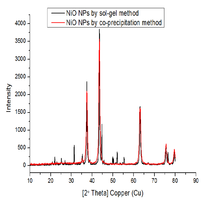
Figure 1: XRD Pattern of NiO NPs Prepared by Co-Precipitation and Sol Gel Methods
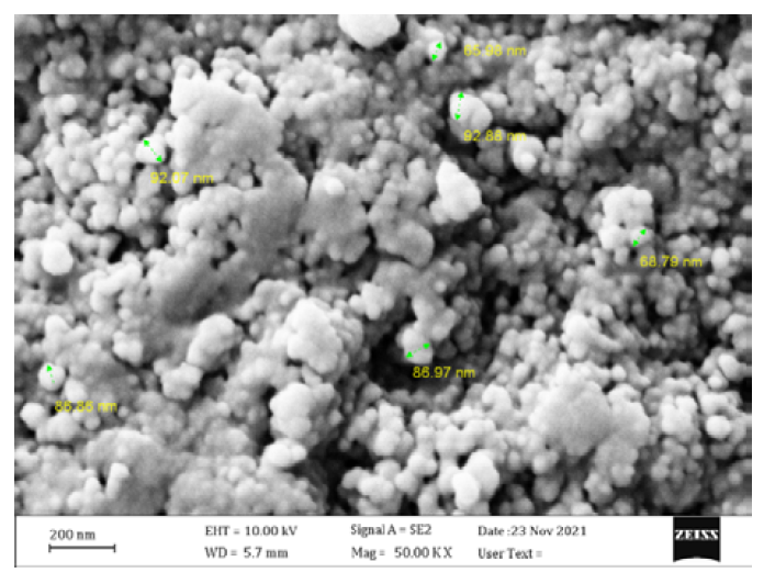
Figure 2: FE-SEM Image of NiO NPs Synthezied by Co-Precipitation Method
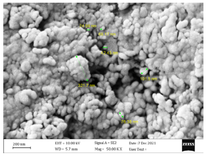
Figure 3: FE-SEM Image of NiO NPs Synthesized by Sol Gel Method
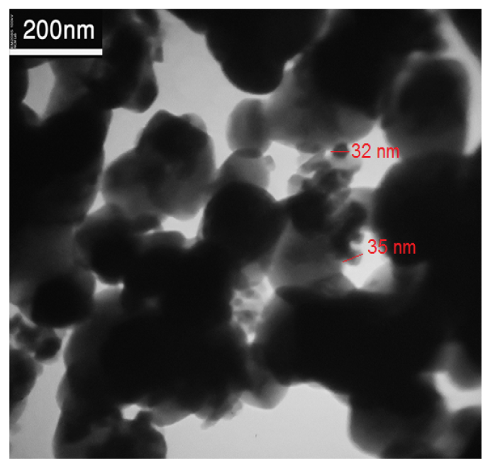
Figure 4: TEM Image of NiO NPs Synthezied by Co-Precipitation Method
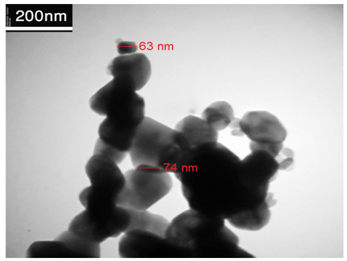
Figure 5: TEM Image of NiO NPs Synthezied by Sol Gel Method
FE-SEM images
Field emission scanning electron microscopy is used to examine the surface morphology of the produced NiO nanoparticle. The co-precipitation method's produced NiO nanoparticles are seen in Figure 2 FE-SEM image. It demonstrates how the particles aggregate to form formations resembling islands, despite their somewhat asymmetrical shapes. An in-depth examination of the island diameters revealed an average diameter of 81.5 nm. Field emission scanning electron microscopy is used to examine the surface morphology of the produced NiO nanoparticle. The FE-SEM image presented in Figure 3 illustrate the uneven aggregation of NiO nanoparticles with an average diameter of 98.66 nm obtained using the sol-gel technique of NiO synthesis.
Figures 2 and 3 may be compared to see how the nickel oxide nanoparticles are different. It is evident that the majority of the nickel oxide appears to be entirely agglutinated using the sol-gel method. One possible explanation for the dispersion of the NiO nanoparticles might be because they are distributed throughout the NaOH solution structure, which is a feature shared by all metal oxides synthesized by the co-precipitation method.
TEM image
TEM is frequently used to image and analyze nanoparticles to determine their size, shape, and morphology. Figures 4 and 5 depict TEM images of the as-prepared, spherically-shaped NiO nanoparticles produced via sol gel and co-precipitation. There are some aggregated particles in addition to the individual ones. For NiO NPs produced via co-precipitation and sol gel, respectively, the detected particle sizes fall between 30 and 35 nm and 65 and 75 nm, respectively.
Antibacterial activity
The antibacterial activity of the produced nanoparticles, NiO NPs, against E. coli and Staphylococcus aureus at concentrations of 100 and 200 mg/mL was assessed using co-precipitation and sol-gel routes. The findings are shown in Table 1. Table 1 shows that the test chemicals' antibacterial effectiveness ranged from good to outstanding. In the two concentrations of 100 and 200 mg/mL, the produced compounds exhibit activity against Staphylococcus aureus that is almost identical to that of the reference. At 100 and 200 mg/mL, the activity against E. coli was outstanding. Due to the drug's resistance to its somewhat effective effects on them.
Table 1: Antibacterial Activity Against E. coli and Staphylococcus Aureus (100 and 200 mg/mL)
| Sample | S. Aureus | E. coli | ||
100 | 200 | 100 | 200 | |
NiO by co-precipitation | 15 | 31 | 24 | 25 |
NiO by sol-gel | 27 | 42 | 23 | 28 |
Ampicillin | 22 | 23 | R | R |
DMSO | 0 | 0 | 0 | 0 |
NiO nanoparticles have been effectively produced using the co-precipitation and sol gel techniques. According to XRD, the synthesis of NiO nanoparticles produced average particle sizes of 34.92 nm for sol gel with a lattice parameter (a = b = c) of a = 4.1786 Å and 27.13 nm for co-precipitation techniques of NiO nanoparticles with a = 4.1684 Å. Although the particles have somewhat asymmetrical shapes, with an average size diameter of 81.5 nm for the co-precipitation method, the FE-SEM image of the NiO nanoparticles shows how the particles aggregate to form formations resembling islands. On the other hand, the image of the NiO produced by sol gel illustrates the uneven aggregation of NiO nanoparticles, with an average diameter of 98.66 nm. The TEM pictures of the spherically-shaped, as-prepared NiO nanoparticles made via co-precipitation and sol gel. In addition to the individual particles, there are also some aggregated ones. The observed particle sizes for NiO NPs generated via sol gel and co-precipitation are 65 and 75 nm and 30 and 35 nm, respectively.
Bayda, S. et al. “The history of nanoscience and nanotechnology: from chemical–physical applications to nanomedicine.” Molecules vol. 25, no. 1, 2020, p. 112.
Bayda, S. et al. “The history of nanoscience and nanotechnology: from chemical–physical applications to nanomedicine.” Molecules vol. 25, no. 1, 2020, p. 112.
Khan, Y. et al. “Classification, synthetic, and characterization approaches to nanoparticles, and their applications in various fields of nanotechnology: a review.” Catalystsvol. 12, no. 11, 2022, p. 1386.
Ju, Y. et al. “Nanoparticles in the earth surface systems and their effects on the environment and resource.” Gondwana Research vol. 110, 2022, pp. 370–392.
Saleh, H.M. and Hassan, A.I. “Synthesis and characterization of nanomaterials for application in cost-effective electrochemical devices.” Sustainability vol. 15, no. 14, 2023, p. 10891.
Khan, I. et al. “Nanoparticles: properties, applications and toxicities.” Arabian Journal of Chemistry vol. 12, no. 7, 2019, pp. 908–931.
Joseph, T.M. et al. “Nanoparticles: taking a unique position in medicine.” Nanomaterials vol. 13, no. 3, 2023, p. 574.
Nikolova, M.P. and Chavali, M.S. “Metal oxide nanoparticles as biomedical materials.” Biomimetics vol. 5, no. 2, 2020, p. 27.
Muscetti, O. et al. “Intracellular localization during blood–brain barrier crossing influences extracellular release and uptake of fluorescent nanoprobes.” Nanomaterialsvol. 13, no. 13, 2023, p. 1999.
Nikolova, M.P. and Chavali, M.S. “Metal oxide nanoparticles as biomedical materials.” Biomimetics vol. 5, no. 2, 2020, p. 27.
Szczyglewska, P. et al. “Nanotechnology–general aspects: a chemical reduction approach to the synthesis of nanoparticles.” Molecules vol. 28, no. 13, 2023, p. 4932.
Wieszczycka, K. et al. “Surface functionalization: the way for advanced applications of smart materials.” Coordination Chemistry Reviews vol. 436, 2021, p. 213846.
Negrescu, A.M. et al. “Metal oxide nanoparticles: review of synthesis, characterization and biological effects.” Journal of Functional Biomaterials vol. 13, no. 4, 2022, p. 274.
Tiwari, M. et al. “0D, 1D, and 2D magnetic nanostructures: classification and their applications in modern biosensors.” Talanta Open vol. 8, 2023, p. 100257.
Byakodi, M. et al. “Emerging 0D, 1D, 2D, and 3D nanostructures for efficient point-of-care biosensing.” Biosensors and Bioelectronics: X vol. 12, 2022, p. 100284.
Joudeh, N. and Linke, D. “Nanoparticle classification, physicochemical properties, characterization, and applications: a comprehensive review for biologists.” Journal of Nanobiotechnology vol. 20, 2022, p. 262.
Abu-Dief, A.M. “Development of metal oxide nanoparticles as semiconductors.” Journal of Nanotechnology and Nanomaterials vol. 1, no. 1, 2020, pp. 5–10.
Deka, B. et al. “Biological and non-conventional synthesis of zinc oxide nanoparticles (ZnO-NPs): their potential applications.” Journal of Nanotechnology and Nanomaterials vol. 3, no. 2, 2022, pp. 79–89.
Pooyandeh, S. et al. “In situ deposition of NiO nanoparticles on cotton fabric using sol–gel method: photocatalytic activation properties.” Journal of Materials Research and Technology vol. 12, 2021, pp. 1–14.
