+91 6002993949
submission@iarconsortium.org
Open Access
ISSN (Print) : 2788-8843
ISSN (Online) : 2788-8851
Dyes are referred to as stains which are used to add color to tissues and microbes to make them optical distinct. The dyes are both synthetic and natural, the synthetic ones may be harmful, hazardous to health and are costly to obtain. Natural dyes are eco-friendly, not costly and not hazardous to health as compared to synthetic dyes. In this study, various procedures such as H&E which serves as control as normal routine staining in histopathology laboratory was used to compare Beetroot and Eosin (B&E), Haematoxylin and Turmeric (H&T). Beetroot and Turmeric (B&T) Beetroot and Eosin stains collagen fibre almost similar to H&E, that shows beetroot can be used as alternative for Haematoxylin in H&E staining technique. Haematoxylin and Turmeric stained the tissue almost similar to H&E which shows that Turmeric can replace Eosin. However, B&T poorly stained collagen fibre when compared with H &E. The conclusion came out that, Beetroot containing Betanin which gives it a red color has the same ability of staining Nuclei Bluish because of its basic content and may require a mordant (Vineger) as attain enhancer for most of the tissues. Turmeric exhibits the basophilic-like effect on collage fibre, it stains the cytoplasm fairly yellowish, because of its curcumin that makes it yellow Beetroot and Turmeric as a direct replacement of H&E staining technique in Histopathology laboratory should further be studied with adequate concentration and mordant as the features is suggestive that B&T can replace H&E.
Dyes are referred to as stains which are used to add to tissues and microbes to make them optical distinct [1]. Stain is a coloration that can be clearly distinguished from the surface, material, or medium it us found upon. They are generally used to add color to plant and animal tissues, microbes and spores to make them optically distinct.
Since immemorial time, natural colors are of paramount importance in dyestuff products but have drastically disappeared owing to the evolution of synthetic dyes. The exploitation of colors cannot be halted, because consumers always demand colored products for eye-appeal, decoration & for aesthetic purposes. Synthetic dyes used in Medical Laboratories such as Hematoxylin and Eosin (H&E) could be replaced with Beetroot and Turmeric (B&T) as their alternative dyes for Histological staining.
Hematoxylin and Eosin (H&E) is a globally practiced staining technique for Histology and Histopathology studies. H&E stains have been used for at least a century and are still essential for recognizing various tissue types and the morphological changes that form the basis of contemporary Cancer diagnosis. In a typical tissue, nuclei are stained blue by the basic dye hematoxylin, whereas the cytoplasm and extracellular matrix have varying degrees of pink staining with acidic Eosin [2].
Many developing countries can no longer afford the ever-increasing cost of synthetic dyes, the use of cheaper, naturally occurring dyes from plants is been viewed as an alternative to synthetic dyes. Based on the fact that, Turmeric (Curcuma long a) was investigated as a natural dye with potential Histopathological application [3].
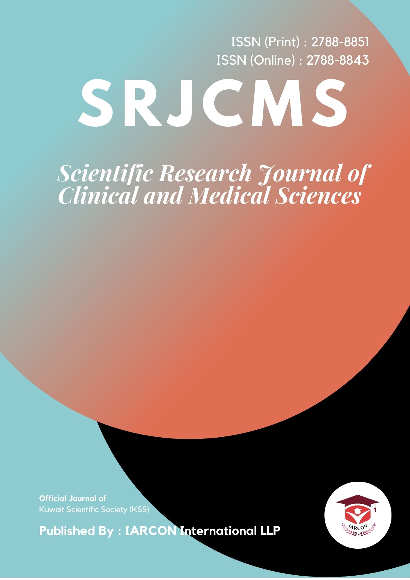
Turmeric (Curcuma longa Linn) has a major component known as Curcumin. It has been consumed as a dietary spice. Chemically, Curcumin is a diarylheptanoid, belonging to the group of curcuminoids, which are natural phenols responsible for Turmeric's yellow color. It contains flavonoids, which are typically polyphenolic compounds. Phenols are acidic, due to their ability to release the hydrogen from their hydroxyl group, hence the ability of Curcuma longa to stain the basic parts of the cell. Curcuma longa was used as a counter stain for Hematoxylin with the reaction like that of Eosin in the Hematoxylin and Eosin technique except for its yellow coloration.
Beetroot (Beta vulgaris) belongs to the chenopodiaceae family [4]. The main pigment found in Beetroot is recognized as Betalain and it is one of the richest source of Betalain which is used for imparting a desirable red color [5]. Beetroot is cherished for its nutritional and Medicinal values thus, it is used in cuisines, salads and juices. It produces purple to red color. Thus, beetroot extract could be used as dye in place of Hematoxylin because, they are all basic dyes and can stain acidic parts of the cell eg nuclei. There are several natural products such as Henna, Blackcurrant, Surreal, kola nut and ginger. For this study, is Beetroot and Turmeric only.
The research work done by Rubina, et al. [6] in Histopathology Laboratory of a tertiary Health care center in South India with "Assessment of staining quality of Curcumin as a substitute for Eosin in Hematoxylin and Eosin staining in Histopathology" concluded that Hematoxylin and Curcuma longa Linn (Curcumin) staining gave a comparable results with Hematoxylin and Eosin with intense affinity on collagen and muscles fibres. They concluded that Curcumin is a safer and cheaper alternating dye (stain) to Eosin stain in Histopathology sections. (H & E) could be (H & T).
Beetroot (Beta vulgaris) was used in Cytology for staining of buccal smears. Beetroot dye (stain) was applied as a fluorescent dye in tissue staining [7]. Researchers such as Avwioro, et al. [8], Bassey, et al, [9]. Conducted the study on soxhlet extracts of Turmeric. They found that, the Turmeric dye stains collagen and muscle fibres with deep yellowish orange color suggesting it's stronger affinity to these structures. A study conducted by Avwioro, et al. and Bassey, et al. also discovered that shelf life of Turmeric was found to be inferior to Eosin because, Turmeric is a natural dye so, when stored for a long period of time, it looses it's color. Kumar, et al. [3] stated that, addition of a mordant to Turmeric can increase the shelf life of this stain.
Recently, Singnarpi have applied beetroot in Cytology for staining of buccal smears and Das [7] have applied beetroot dye as a fluorescent dye in tissue staining properties when compared with Hematoxylin and Eosin (H & E) counterparts.
Hematoxylin is a basic dye which stains the nucleus and some parts of cytoplasm containing the nucleic acid or acidic structures blueish purple or black in some instance. Haematoxylin and Beetroot extract have similar staining effects staining tissue. Beetroot extract exhibited Haematoxylin like staining effect. On the contrary, Eosin, an acidic dye will stain basic structures deep pink color. The exceptions to these are neutral cellular and extracellular components that take up neither of these and appear relatively clear [10]. This implies that, the extracts having exhibited Haematoxylin property, Beetroot extract can be used in combination with Eosin (B&E), in place of (H& E).
The bright yellow colored compound found in Turmeric is called Curcumin. Such main fluorescent component or the Curcumin in Tumeric is so dominant that it could be used as a stain. The study conducted by Kumar [3] revealed that, natural tint from Turmeric could stain tissues such as collage and muscle fibers.
The Curcumin in Turmeric which gives a yellowish crystalline color is acidic dye such as Eosin in Histopathology Laboratory.
Eosin is an acidic dye (stain) used as a counterstain in Haematoxylin and Eosin routine technique. It stains the basic parts of the cells such as cytoplasm deep pink color.
Turmeric Extract could stain tissues such as collage muscle fibers [3]. Turmeric has a good potential and promising histological dye that can excellently replaced Eosin stain in Haematoxylin and Eosin routine. In my study, it could be Haematoxylin and Turmeric (H&T).
The aim of this research work is to prove that natural products Beetroot and Turmeric can be used as alternative dyes for Hematoxylin and Eosin in Histopathology Laboratories with specific objectives:
To describe how turmeric can replace eosin in histological staining.
To describe how beetroot can replace haematoxylin in histological staining.
To educate Nigerians and researchers that local products like beetroot and turmeric can replace haematoxylin and eosin in histology.
Collection of both Turmeric and Beetroot Products
Turmeric and Beetroot plants are cultivated in Plateau State, Jos Nigeria. The products were bought from Terminus Market, Jos-Nigeria.
Preparation of natural dye materials
10g of Beetroot bulb and 10g of Turmeric rhizome were washed with clean water, after removing the soil. The outer parts were removed, and blended using blender. Each stain was dissolved in 1L of distilled water and were collected through sieving with a clean sieve clothe
material, the extracts were allowed to settle in to test tubes and the supernatants were kept in to the refrigerator at 4°c. The extracts were ready for use afterfiltration using Wattman No 1 filter paper in to separate staining bottles from labelled concentrated beetroot and turmeric extracts. as shown in Figures 3 and 4.
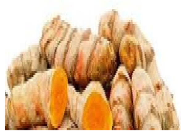
Figure 1: Turmeric roots (Fresh)
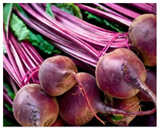
Figure 2: Beetroot (Fresh)
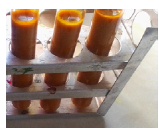
Figure 3: Turmeric extract.
Preparation of Haematoxylin and Eosin
The Haematoxylin and Eosin were prepared according to Ochei and Kolhatkar,[2].
Staining of Different Tissue Sections
Collagen tissues were stained using both Beetroot and Turmeric extracts while Haematoxylin and Eosin served as control stains.
Procedure for Haematoxylin and Eosin staining Technique. (H&E)
The procedure was in line with Ochei and Kolhatkar, [2] using the steps as below:
Take section to water.
Stain section in Haematoxylin solution for 5mins.
Rinse in water for few seconds.
Differentiate in 1% acid alcohol with continuous agitation for 10 seconds.
Blue in Scott tap water for 5mins.
Counter stain in 1% aqueous Eosin solution for 5mins.
Wash in running tap water for 30secs.
Dehydrate in ascending grades of alcohol.
Clear in xylene
Mount in DPX.
Procedure for Staining Tissue with Beetroot and Turmeric (B&T)
The steps adopted for the staining is as shown below:
Take section to water.
Stain with beetroot for 10mins.
Wash with vinegar.
Differentiate with 1% acid alcohol for few seconds.
Wash in water.
Blue with Scott's tap water for few seconds.
Wash with water.
Counterstain with Turmeric for 5 mins.
Wash with water.
Dehydrate with ascending grades of alcohol.
Clear with xylene.
Mount with DPX and coverslip.
Procedure for Haematoxylin and Turmeric (H&T).
Take section to water.
Stain section in Haematoxylin for 5 mins.
Wash in water.
Differentiate in 1% acid alcohol for few seconds.
Wash in water.
Blue in Scott's tap water briefly.
Wash in water.
Counterstain in Turmeric for 5 mins.
Wash in water.
Dehydrate in ascending grades of alcohol for 2 mins.
Clear in xylene.
Mount with DPX and cover with coverslip.
Procedure for Beetroot and Eosin (B&E)
Take section to water.
Stain in beetroot for 10 minutes
Wash in vinegar.
Differentiate in 1% acid alcohol for few seconds.
Wash in water.
Blue in Scott's tap water briefly.
Wash in water
Counterstain in 1% aqueous Eosin solution for 5 minutes
Wash in water.
Dehydrate in ascending grades of alcohol for 2 minutes.
Clear in xylene
Mount with DPX and cover with coverslip.
Haematoxylin and Eosin serve as a control; it stains collagen fibre. The nuclei-stained bluish purple with Haematoxylin and cytoplasm-stained pink with Eosin.
Haematoxylin and Turmeric used on collagen fibre. Nuclei-stained bluish purple with Haematoxylin and cytoplasm stained yellowish with Turmeric.
Beetroot and Eosin is used on collagen fibre. Nuclei stained Blusish purple with Beetroot and cytoplasm-stained pink with Eosin.
Beetroot & Turmeric used on collagen fibre. Nuclei stained Black Beetroot and cytoplasm stained yellow with turmeric.
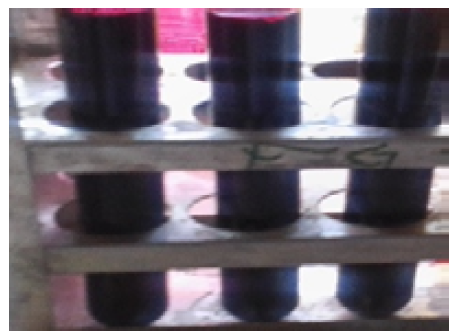
Figure 4: Beetroot extract.
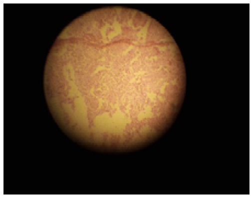
Figure 5: Collagen Fibre stained with Hematoxylin and Eosin (Mx100).
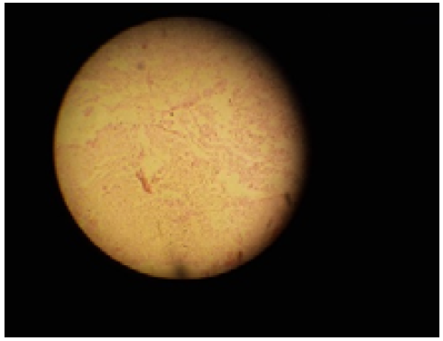
Figure 6: Collagen Fibre stained with Hematoxylin and Turmeric (Mx100).
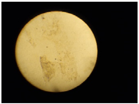
Figure 7: Collagen Fibre stained with Beetroot and Eosin (Mx100).
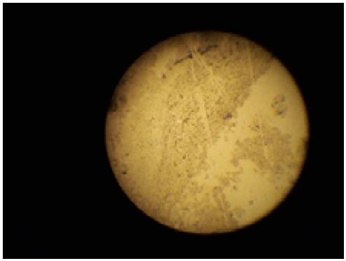
Figure 8: Collagen Fibre stained with Beetrrot and Turmeric (Mx100).
According to the results shown in the pictures below, (H & E) stains Nuclei blue purple and cytoplasm pink. (H and T) Stain Nuclei blue-purple and cytoplasm yellowish. (B & E) stains nuclei bluish purple and cytoplasm pink. (B & T) stains nuclei bluish purple and cytoplasm yellowish. The study shows some resemblance and unique features on the cells and cytoplasm which are directly noted in the tissue except for H&T which is different from H&E. this may be due to colours, concentration, and mordant.
This suggests that beetroot extract exhibited heamatoxylin like staining effects. Eosin as an acidic dye will stain basic structures pink colour. This implies that, the Beetroot extract having exhibited Haematoxylin property, it can be used in combination with Eosin, (B & E) place of (H & E) [10]. On the other hand, acidic dye which stains basic structures red or pink colour. Turmeric extract as a natural dye exhibited like staining property of Eosin as having acidic content to stain basic structures yellowish colour because of its curcumin (Fiqure 8). This suggest that because of the acidic content of turmeric like Eosin, it can be used inplace of Eosin in H & E staining technique as H & T. But because of yellowish coloreation and poorly stained slides, further studies should be carried out on Beetroot and turmeric.
Heamotoxylin and Eosin are the most considerable stains used in Histological staining. In this study Beetroot & Turmeric extract are used for staining collagen fibres. Beetroot adequately replaced Hematoxylin and Turmeric adequately replaced Eosin in Hematoxylin and Eosin methodology. However, Beetroot and Turmeric could not adequately replace Hematoxylin and Eosin and it is recommended that more studies should be carried out based on the concentrations and adequate mordants to use for adequate replacement of Hematoxylin and Eosin (H&E) by Beetroot and Turmeric (B&T).
Ragaswami, G., et al. Agricultural Microbiology. 2nd ed., Practice-Hall of India Private Limited, 1993, pp. 18–29.
Ochei, J., and J. Kolhatkar. Medical Laboratory Science: Theory & Practice. 3rd reprint, 2007, p. 441.
Kumar, N., et al. “Staining of Platyhelminthes by Herbal Dyes: An Ecofriendly Technique for the Taxonomist.” Veterinary World, vol. 8, 2015, pp. 1321–1325.
Prabhu, K.H. “Plant-Based Natural Dyes and Mordants: A Review.” Journal of Natural Product and Plant Resources, vol. 2, no. 6, 2012, pp. 649–664.
Gaszt́onyi, M.N., et al. “Comparison of Red Beet (Beta vulgaris var. conditiva) Varieties on the Basis of Their Pigment Components.” Journal of the Science of Food and Agriculture, 2001.
Rubina, M.P., et al. “Assessment of Staining Quality of Curcumin as a Substitute for Eosin in Hematoxylin and Eosin Staining in Histopathology.” Journal of Research in Medical and Dental Science, vol. 8, no. 5, 2020, pp. 146–150.
Das, A., et al. “Improvement in Cell Imaging by Applying a New Natural Dye from Beetroot Extraction.” arXiv, 2017, https://arxiv.org/pdf/1710.08119.pdf.
Avwioro, O.G., et al. “Extracts of Pterocarpus osun as a Histological Stain for Collagen Fibers.” African Journal of Biotechnology, 2005.
Bassey, R., et al. “Curcuma longa Linn: Staining Effects on Histological Morphology of the Testis.” Macedonian Journal of Medical Sciences, 2012.
Anneh, H., et al. “Natural Dye for Staining Astrocytes and Neurons.” Journal of Neurological Sciences, 2006.
