
+91 6002993949
submission@iarconsortium.org
Open Access
ISSN (Print) : 2709-3239
ISSN (Online) : 2709-3220
Head injury occurs as a result of trauma to the scalp, skull/ brain. It may be closed where there is no cut to the skin or penetrating (skin/bone of the skull is broken). India has an unenviable distinction in having Highest rate of death due to injury in the world. More than 100,000 lives are lost every year with over one million suffering from serious head injuries. Sub-arachnoid haemorrhage is most common in Traumatic Brain Injury.
Head injury is mainly caused during fall from a height, road traffic accidents and assaults targeting head, which was the case here. In case of assaults, using blunt weapons, head is the soft and most targeted organ as the as it proves to be lethal because of the injuries caused to brain and other associated delicate organs. In India, 1 out of 6 trauma victims die due to head injury. The death is within two hours of injury in 50% of the victims. It is because at the time of impact, the neurological damage is partial sometimes (primary), which progresses further (secondary), worsening the situation, resulting in death [1,2].
A tea-stall owner inside the BHU campus was brutally murdered by robbers who attacked him early in the morning while in sleep. The weapons used for the purpose were brick and large stone pieces present at the scene of occurrence itself. The body part targeted was head using the mentioned weapons resulting in blunt trauma to the different part of skull and brain.
This article is about the penetrating head injury, where the death was caused on the spot as a result of coma due to haemorrhage and excessive bleeding.
The head injuries included:
Concussion-including injuries to the brain caused by blow to the head or body, shaking the brain inside the skull.
Contusion-bruising of brain tissue associated with swelling edema and intracranial pressure.
Fracture-crack or break in the skull, with laceration to the skin. The fracture encountered in the above case was depressed signature fracture on the forehead and depressed comminuted fracture present on the skull.
Bleeding-owing to the hemorrhage due to bursting of blood vessels. Blood may had collected and formed hematoma i.e, epidural (clotting between the inside of the skull and dura matter) and sub-dural hematoma (blood collecting beneath dura matter).
Shear injury-also called diffuse axonal injury. Brain bounces violently against the inside of the skull (Head Injury. (2019).
Skull Fracture
Fractures are described based on location, as all the skull bones are not the same in fragility. Depending upon the fracture location, there may/may not be associated brain injury.
The above case included:
Basilar- because of blunt trauma and damage to the base of the skull, especially anterior cranial fossa, resulting in racoon eyes .
Depressed skull fracture- The piece of skull was pushed toward the inside of the skull, inner table of the skull bone is fractured.
Bleeding (Intra-cranial)
Sub-dural Hematoma-occurring as a result of tearing and bleeding of bridging veins that cross through the subdural space, as a result of the applied force. It may sometimes occur as the site of the trauma or on opposite side of the injury (contra-coup).
Epidural hematoma as a result of trauma to the temporal bone located on the side of the head above the ear. The fracture of the temporal bone is associated with the tearing of the middle meningeal artery, running just beneath it which led to epidural hematoma.
Sub-arachnoid hemorrhage-blood accumulated in the space beneath inner arachnoid layer of the meninges associated with intra-cerebral bleed, mainly due to rupture of anterior communicating artery.
Intra-cerebral haemorrhage-bleeding within brain tissues itself [1].
The above case was brought to autopsy room of Sir Sundar Lal hospital, Institute of Medical Sciences, Banaras Hindu University. The case was studied carefully and injuries were noted. The photographs were taken at the time of post-mortem and were studied for the inference of injuries present.
A detailed search was made through the web from Google scholar, web med and pub med, searching the keywords-Head injury and associated haemorrhage, sub-arachnoid haemorrhage, temporal bone fracture and incidence of fracture in head trauma. The materials were thoroughly studied and the material relevant to the case was sorted out.
Comparative study was made with the relevant literature material collected from different sources.
Figure 1 depicted blackening around the eyes also called “Racoon eye”. Medically also known as periorbital ecchymosis, it is bruising and discolouration around the eye. The most common cause and also the cause of Racoon’s eye in the above case is injury to the head, leading to Basal Skull Fracture, including temporal bone, parietal bone or ethmoid bone. The other cause may be facial fractures, involving the fracture of thin bones surrounding the eyes. Further, broken nose, cheek bone, and broken eye socket may be the possible reasons for the same [3].
Figure 1: Photograph Depicting Black Eye
The incidence of racoon eyes associated with Skull base fracture is 50-60%, which can be attributed to anterior skull base fractures, especially of the frontal bone with associated epidural hematoma [4].
In the above case unilateral racoon eye is evident, which is indicative of basilar skull fracture. The incidence of unilateral Racoon Eyes Sign is 74% and bilateral RES is 77% [5].
Figure 2 showed nasal bleeding which can be attributed to head injury, which may result in bleeding around the brain [6]. Olfactory nerves being closely connected with ethmoid bone, has numerous nerves passing through cribriform plate of the ethmoid bone, which gets damaged as a result of assault or trauma to the anterior cranial fossa, of which it forms a part, resulting in nasal bleeding [7].
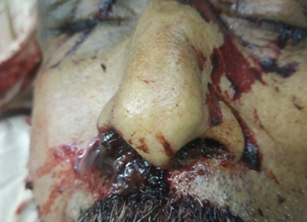
Figure 2: Bleeding from the Nostrils as a Result of Fracture to the Base of Brain
Figure 3Bleeding from the ear may be attributed to head injury. The damage to the temporal bone can be caused because of blow to the head, causing damage to the middle cranial fossa which may result into bleeding from the ear [8]. If bleeding from the ear accompanies head injury, it may be due to concussion [9].
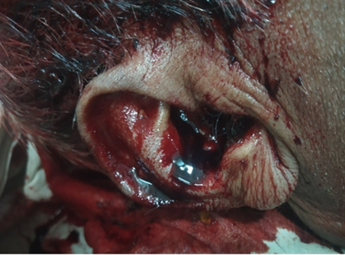
Figure 3: Bleeding from Ear as a Result of Fracture of Base of the Brain
Figure 4 showed signature fracture. Fracture-a-la-signature is another term used to describe depressed skull fracture. The size and shape of a depressed skull fracture may be informative about the type of weapon used. I n the above case, the weapon used for assault was corner of the brick which can be co-related with the fracture produced [10].
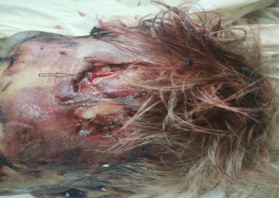
Figure 4: Photograph depicting signature fracture
Figure 5 depicts the fracture in the temporal bone are lesions, observed in patients with traumatic brain injury (TBI).It is frequently associated with severe brain injury. Fracture is evident in approximately 4% of the patients of head injury. Out of these 14-22% presents fracture of the temporal bones. The foremost cause is vehicular accidents (45%), falls (31%) and robberies, which was the cause in above case. The fracture of temporal bone is associated with further complications leading to hearing loss, facial paralysis and otogenic CSF leakage.
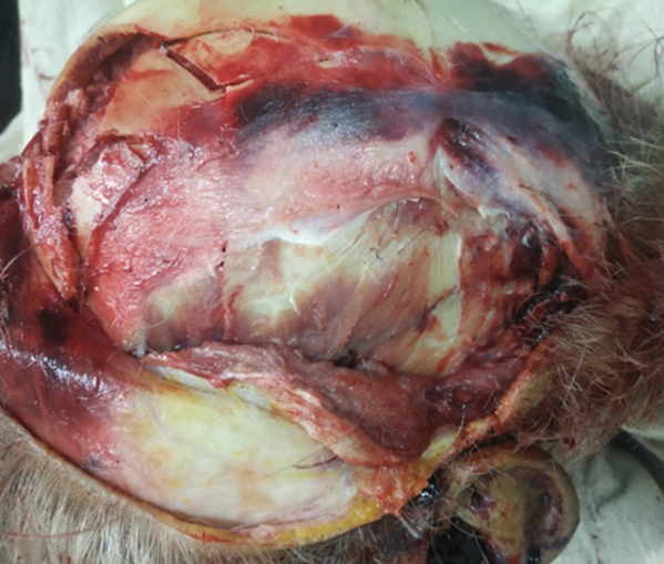
Figure 5: Photograph Depicting Fracture Of Temporal Area With Laceration Of Dura Matter
Temporal bone in the skull is the common bone to fracture in head injuries as it is very thin, with a thickness of 4mm.
Further, the damage to the top floor of the middle fossa and the region of middle ear and mastoid and its disruption can produce a fistula, leading to leakage of CSF, if tympanic membrane remains intact [11].
Dural tear is usually caused as a result of trauma or as a complication of surgery. In the above case i.e, the case of assault involving head injury, dural tear is usually seen in case of depressed skull fracture [12].
Figure 6 showed the depressed comminuted fracture. According to Aggarwal A, the fracture is also known as Spider’s web or mosaic fracture. In this kind of fracture there are two or more intersecting lines which divide the bone into three or more fragments. Since the bone fragments have been pushed inwards after the fracture, in the above case, it is depressed comminuted fracture. The cause of this fracture is fall from height on a hard surface, animal kicks, vehicular accidents and weapons with large striking surface, the last one being the cause in this case. The fracture lines are defined by Puppe’s rule [13].
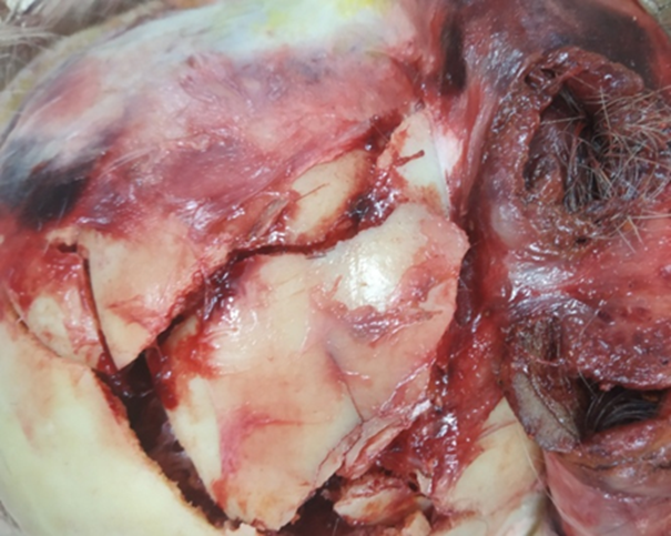
Figure 6: Depressed Comminuted Fracture In The Fronto-Parietal Area Due To Heavy Impact
Figure 7 It showed sub-arachnoid haemorrhage. Sub-arachnoid haemorrhage refers to extra vasation of blood into the sub-arachnoid space between the pial and arachnoid membrane as a result of head trauma in this case [11]. It is the most common CT finding in TBI. Sub-dural and epidural hematoma are the most frequent type of lesion found in TBI [14].
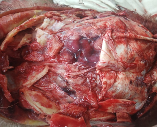
Figure 7: On Opening The Skull, Laceration Of Dura Matter Was Seen Along With Sub Arachnoid Haemorrhage
Figure 8 depicted cadaveric spasm. According to Aggarwal A, it is also known as instant rigor, post-mortem spasm or cataleptic rigidity. It is a rare condition, in which the muscles that were in contraction at the moment of death, remain in contraction after death without passing through the stage of primary relaxation. The predisposing condition relevant to this case may be cerebral haemorrhage. One of the reasons is attributed to ATP depletion at the time of death due to muscles in action, which in this case may be due to the resistance offered. This is why the last activity of the person has frozen for hours after the death [13].
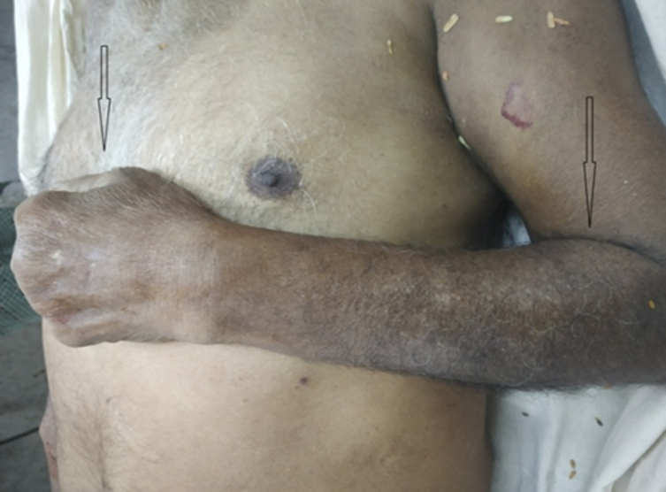
Figure 8: Cadaveric Spasm Showing That Muscle Was In Action At The Time Of Death
The above case presented the homicidal head injuries which proved to be fatal to the victim. The injuries to the head led to fractures in the skull along with associated brain injuries causing bleeding and finally death as a result of coma. The further complications could not be studied as the death occurred immediately after the incident on the spot itself.
Authors’ Contribution
Both the authors have contributed in post-mortem, analysing the photographs of the case and preparation of the manuscript.
Traumatic Brain Injury. Indian Head Foundation, August 2021, https://indianheadinjuryfoundation.org/traumatic-brain-injury/.
Head Injury. RadiologyInfo.org, January 2019, file:///C:/Users/Dell%20Pc/Downloads/headInjury%20(2).pdf. pp. 2–3.
Johnson, J. “What’s to Know About Racoon Eyes.” Medical News Today, August 2017, https://www.medicalnewstoday.com/articles/319039#causes.
Das, J.M., and S. Munakomi. “Racoon Sign.” StatPearls, February 2021, https://www.ncbi.nlm.nih.gov/books/NBK542227/.
Herbella, F.A.M., et al. “ ‘Racoon Eyes’ (Periorbital Hematoma) as a Sign of Skull Base Fracture.” Injury, vol. 32, 2001, pp. 745–747. https://doi.org/10.1016/S0020-1383(01)00144-9.
Holland, K. “What Causes Ear Bleeding.” Healthline, March 2019, https://www.healthline.com/health/ear-bleeding#call-a-doctor.
Adams, B. “The Anterior Cranial Fossa.” TeachMeAnatomy, September 2019, https://teachmeanatomy.info/head/areas/cranial-fossa/anterior/.
Most, S.P. “Temporal Bone Fracture.” MSD Manual, Consumer Version, May 2020, https://www.msdmanuals.com/en-in/home/injuries-and-poisoning/facial-injuries/temporal-bone-fracture.
Fletcher, J. “Bleeding from the Ear: Causes and Treatments.” Medical News Today, December 2017, https://www.medicalnewstoday.com/articles/320237#anatomy-of-the-ear.
Bell, D.J., and D.H. Knipe. “Fracture-a-la-Signature (Skull Fracture).” Radiopaedia, 2021, https://radiopaedia.org/articles/fracture-a-la-signature-skull-fracture.
Secchi, M.M.D., et al. “Fracture of the Temporal Bone in Patients with Traumatic Brain Injury.” Arquivos de ORL, 2012, http://arquivosdeorl.org.br/additional/acervo_eng.asp?id=829.
“Dural Tear.” Wikipedia, November 2019, https://en.wikipedia.org/wiki/Dural_tear.
Aggarwal, A. Textbook of Forensic Medicine and Toxicology. Avichal Publishing Company, 2017. pp. 327.
Becske, T. “Sub-arachnoid Hemorrhage.” Medscape, December 2018, https://emedicine.medscape.com/article/1164341-overview.
