Malaria infection during pregnancy is an enormous public health problem, with substantial risks for the mother, the feotus and the neonate. The study was carried out in Primary Health Care Antenatal clinic Damaturu in which 200 pregnant were involved between the months of July and September 2015. Peripheral blood samples were collected using venous procedure and the presence of malaria parasites was observed microscopically on thick and thin blood smears prepared from each sample to determine the degree of parasitaemia. 115 (representing 57.5%) pregnant women were found to be infected with malaria parasite. Personal data were collected through questionnaires and the general results obtained during this study were analyzed statistically using simple percentage. Where adequate medical care is available, however, certain antenatal interventions appear to be effective in reducing maternal mortality due to malaria in pregnancy.
Keywords
Malaria
Parasitemia
Pregnancy
INTRODUCTION
Malaria is a parasitic infection caused by a unicellular protozoan parasite of the genus plasmodium [1]. It is transmitted by the bite of female anopheles’ mosquito. It is one of the most devastating infectious diseases, killing more than one million people annually [1]. Pregnant women, children and immuno compromised individuals have the highest morbidity and mortality and Africa bears the heaviest burden [1].
According to Sketekee & Mutabingwa, malaria in pregnant women, [2], malaria is a life-threatening parasitic disease transmitted by female Anopheles mosquitoes and caused by different species of Plasmodium parasites. An increased risk of malaria during pregnancy was observed over 60 years ago by Wickramasariya [3]. The protection of pregnant women living in malaria-endemic countries has been of particular interest to many National Malaria Control Programmes because of their reduced immunity [1]. The recent World Malaria Report indicated that Nigeria accounts for a quarter of all malaria cases in the 45 malaria-endemic countries in Africa, which clearly showed the challenge of malaria in Nigeria [1]. The principal impact of malaria infection is due to the presence of parasites in the placenta causing maternal anaemia (potentially responsible for maternal death when severe) and low birth weight (LBW) [4]. Despite considerable efforts to control malaria, it is still the most prevalent and life-threatening disease in tropical Africa (with pregnant women and children below five years the highest risk groups) [5].
Malaria infection during pregnancy is an enormous public health problem, with substantial risks for the mother, her foetus and the neonate [6]. In areas of low transmission of Plasmodium falciparum, where levels of acquired immunity are low, women are susceptible to episodes of severe malaria, which can result in stillbirths or spontaneous abortion or in the death of the mother [7]. In areas of high transmission, of Plasmodium falciparum, where levels of acquired immunity tend to be high, women are susceptible to asymptomatic infection, which can result in maternal anaemia and placental parasitemia, both of which can subsequently lead to low birth weight [8]. Low birth weight is an important contributor to infant mortality. It has been estimated that malaria during pregnancy is responsible for 5-21% of all low birth weight and contributes to 75,000 to 200,000 infant deaths each year [9]. The world Health Organisation (WHO), currently recommends a package of interventions for controlling malaria during pregnancy in areas with stable (high) transmission of P. falciparum, which includes the use of insecticide-treated-nets (ITN’s), intermittent preventive treatment (IPT) with sulfadoxine pyrimethamine (SP) and effective case management of malaria and Anaemia (Personal communication with Daskum A.M).
In Plasmodium vivax infections in pregnancy, severe malaria is very rare. No intense placental sequestration occurs yet the average reduction in birth weight is roughly 107 g (compared with 170 g in falciparum malaria), [10]. Even in symptomatic cases, promptly treated Plasmodium falciparum or Plasmodium vivax infections increase the risks of having abortions and low birth weight [11]. The risk of infant death is particularly high if maternal malaria occurs during late (that is near-term) pregnancy. Maternal death from haemorrhage at childbirth is correlated with malaria-induced anaemia [12].
Many antenatal interventions have been shown to reduce neonatal morbidity and mortality [13], however, evidence for the effectiveness of antenatal care in reducing maternal mortality (and to a lesser extent, morbidity) is less compelling [14]. It is widely accepted that screening pregnant women to identify those at risk for obstetric complications is not a replacement for skilled care during labor and delivery [14]. More maternal deaths occur in the much larger group of low-risk women. As a result, antenatal care will not necessarily prevent complications from occurring [15]. This was demonstrated in a study in Gambia in the early 1980s in which a relatively high standard of antenatal care was not able to identify the specific risk factors that could predict which women were more likely to experience fatal complications [16]. In addition, those who did experience complications were often located too far from a competent medical facility to receive treatment. As a result, maternal mortality remained extremely high at 2,000 deaths per 100,000 live births [16]. Where adequate medical care is available, however, certain antenatal interventions appear to be effective in reducing adverse maternal outcomes [13,17]. These include the recognition and treatment of hypertensive disease of pregnancy, detection and treatment of asymptomatic bacteriuria and external cephalic version at term (to prevent obstructed labor) [13,17], more controversial are antenatal interventions to prevent maternal anemia and other forms of nutritional supplementation. In addition to the potential for reducing specific causes of maternal morbidity and mortality, antenatal care can also encourage birth preparedness and the use of skilled assistance in labor and delivery [13,17].
MATERIALS AND METHODS
Data Source
For the purpose of this research work, one major source of data was utilized. This includes the primary source data. The primary data was collected through oral interview, questionnaires and laboratory experiment (Figure 1).
Sampling Methods and Techniques for Data Collection
For the purpose of this research study, a simple random sampling was carried out. Three nurses at the antenatal clinics located at primary health care and management board were interviewed to identify the number of cases of maternal mortality during pregnancy due to malaria as well as factors that influences malaria distribution during pregnancy.
Similarly, a simple random sampling was used to administer the questionnaires where ten questionnaires were administered to ten pregnant women in each week for a period of ten weeks.
Figure 1: A Map Showing Damaturu Local Government Area, Yobe State
Limitation of Data of Collection
However, there are some problems encountered in caring out the research work which includes lack of adequate fund, time limitation which hindered the interview of some part of the population, poor responses from those interviewed. Lack of understanding particularly from those with low level of western education, socio cultural religious impact particularly from the married women.
Experimental Procedure
The blood sample was collected from 200 pregnant women attending primary health care and management board located at Gwange ward Damaturu Yobe State. Twenty samples were randomly selected and tested for malaria each week to determine the prevalence rate of malaria among pregnant women. The blood sample was collected using the finger pricking method or venous collection.
Pricking Method for Blood Collection
The patient is asked to seat on an appropriate seat, wear a hand glove and use a cotton wool lightly soaked in alcohol to clean the finger, using firm strokes to remove grease from the ball of the finger. The finger is allowed to air dry. With a sterile lancet, puncture the ball of the finger using a quick rolling action. By applying gentle pressure to the finger, express the first drop of blood and wipe it away with dry cotton wool (making sure that no strands of cotton wool remain on the finger). Collect the blood by applying gentle pressure using a plastic pipette [18].
Venous Collection Method
Wear a hand glove and ask the patient to seat on an appropriate seat
Tie the fore arm of the patient with a tony cate and search for the prominent vein
Using cotton wool, slightly soaked with alcohol, disinfect the area of the vein
Fix, the syringe and needle well and insert it carefully and gently into the vein, making sure the sharp edge of the needle is facing upward
Gently and carefully pull the syringe jet, until the required blood mill is reached
Before removing the syringe, untie the tony cate (to restore circulation)
Remove the syringe from the vein carefully, remove the needle from the syringe chamber and discard it into a safety box
Put the collected blood into the appropriate container, example EDTA (Ethylene diamine tetra acetate) container
The collected sample is transported to the haematology unit for malaria laboratory diagnosis [19]
Preparing Thick and Thin Blood Film for Microscopic Analysis
Thick Blood Firm: A thick blood firm is made by putting a drop of blood in the centre of a glass slide using a plastic pipette. Using a dropper, make a circle in form of coin (as described by Swiss tropical institute practical haematology). The firm is allowed to dry well before staining.
Thin Blood Firm
This is made by putting a drop of blood or two drops of blood at one end of the glass slide. A cover slip is placed at one end of the drop with its edge at 45 degrees. This is carefully and gently pushed forward until the firm is made. The film is allowed to dry very well (Swiss Tropical Institute Basel, April).
Staining Procedure
After the thick and thin blood film have been made and allowed to dry. The slides are dipped into Field Stain A for seconds and the excess stain is drained by touching the corner of the slide against the reagent container. The slides are rinsed with water for five seconds. The slides are again dipped into Field stain B for five seconds and the excess are again drained by tapping it unto the reagent container. This is rinsed with water for 5 seconds. The back of slides is wiped and cleaned with cotton wool and this is allowed to dry for microscopic examination [19].
Microscopic Examination
After the blood is collected, smeared on a slide, stained and dried. One or two drops of oil immersion (cederod oil) is placed on the stained film. This is placed on a microscopic stage and viewed by using X100 objective lens (Swiss Tropical Institute, Basel). The malaria parasite present in the red blood cells, were counted were counted per field view.
Packed Cell Volume (PCV)
This refers to the volume of the blood cells in a given sample of blood. This is done to determine if the patient is anaemic as a result of malaria infection. The following procedure is used:
Fill three-quarter ¾ of a haematocrit capillary tube with the uncoagulated blood in EDTA container. The back of the capillary tube is cleaned with a dry cotton wool.
One end of the capillary tube is sealed with a sealant (e.g. crystacille, clay or Bunsen flame). This is inserted into a micro heamatocrit centrifuge machine.
It is allowed to spin at 10,000 revolutions per minute for five minutes. After five minutes, the machine is stopped, the capillary tube is removed and the result is being interpreted using a haematocrit reader [17].
RESULTS
The Table 1,2 show the result obtained from women attending Gwange Ward Primary Health Care Antenatal Clinics. Based on the research carried out, out of 200 pregnant women examined between the period of June 2015 to September 2015, 115 were diagnosed with malaria, which represent 57.5% of the population.
Prevalence of Anaemia in Pregnant Women Attending Antenatal Clinics
After the investigation of the packed cell volume (PCV) level in the infected pregnant women attending antenatal clinics in Damaturu for anaemic condition, the following result was obtained (Table 3 And Figure 2-5).
Table 1: Infection Rate and Prevalence of Malaria in Pregnant Women
Period of
Sampling
Number of
Sample
Number of
infected
Number of not
Infected
week 1
20
14
6
week 2
20
10
10
week 3
20
13
7
week 4
20
15
5
week 5
20
18
2
week 6
20
3
17
week 7
20
11
9
week 8
20
8
12
week 9
20
14
6
week 10
20
9
11
TOTAL
200
115
85
Table 2: Degree of Infection/Parasitemia in Pregnant Women Attending Primary Health Care Antenatal Clinic
Period of
Sampling
Number of infected
Degree of infection
(+)
(++)
(+++)
week 1
14
3
8
3
week 2
10
4
5
1
week 3
13
7
3
3
week 4
15
4
3
8
week 5
18
8
6
4
week 6
3
2
0
1
week 7
11
5
5
1
week 8
8
1
4
3
week 9
14
6
6
2
week 10
9
1
1
7
TOTAL
115
41
41
33
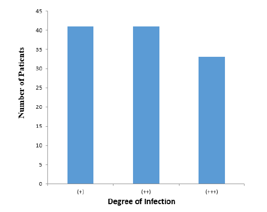
+ = 0ne to ten malaria parasite per field view, ++ = 10-20 malaria parasite per field view, +++ = more than 20 malaria parasite counted per field view
Figure 2: The Bar Chart Above Shows the Various Degree of Infection in Pregnant Women Attending Antenatal Clinics in The Primary Health Care and Management Clinic (Gwange) Damaturu
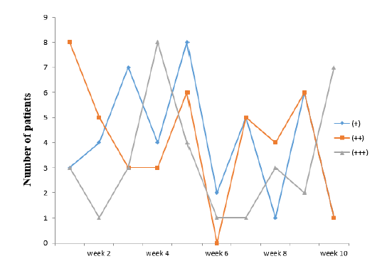
+ = 0ne to ten parasites per field view, ++ = 10-20 parasite per field view, +++ = more than 20 parasite counted per field view
DISCUSSION
Maternal, placental or foetal malaria infection during pregnancy adversely affects pregnant women and the development and survival of foetus through low birth weight, maternal anaemia and possibly abortion and stillbirth [6]. These malarias induced medical problems constitute major clinical, public health and research challenges [20]. In women from non-endemic areas or travellers to endemic areas, malaria infection is associated with high risk of maternal and perinatal mortality [13].
Randomized controlled trial of interventions to reduce malaria in pregnancy has demonstrated some successes in reducing maternal mortality due to malaria in pregnancy through Antenatal Clinics [13]. Furthermore, where adequate medical care is available, certain antenatal interventions appear to be effective in reducing adverse maternal outcomes [17,21]. These include the recognition and treatment of malaria pregnant women, as well as finding ways of preventing its occurrence. In addition to the potential for reducing specific causes of maternal morbidity and mortality, antenatal care can also encourage birth preparedness and the use of skilled assistance in labor and delivery [17].
Table 1 shows prevalence rate of malaria, was high during the period of the research between the months of July to September 2015 as 115 pregnant women were found infected which represent 57.5% of the total pregnant women analyzed. This may be due to high/Frequent exposure of pregnant women to mosquito bites and increasing resistance of the malaria parasites to certain anti-malarial drugs [13].
Table 2 shows the various degree of infection of malaria in pregnant women attending Gwange ward Primary Health Care and management board antenatal clinics Damaturu where 41 are found to show low parasitemia (+), 41 shows a moderate parasitemia (++), while 33 were found to have a high parasitemia (+++). However, (++) and (+++) parasitemia are of concern to pregnant women, as it may lead to maternal anaemia, hygpoglycemia, fever, placental parasitemia, or neonatal death [6].
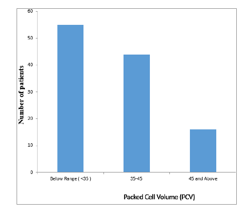
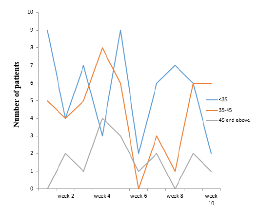
Table 3 shows the packed cell volume (PCV) and the prevalence of anaemia due to malaria parasites infection in pregnant women attending Gwange Ward Primary Health Care Antenatal clinic Damaturu, which shows that: 55 pregnant women were anaemic due to their low PCV level (below 30%) which was accompanied by chills, headache, body pains and sweating. In severe anaemia, the patient complains of extreme fatigue and general body weakness [6]. 44 were not anaemic due to their normal PCV level (35-45%), which is in agreement with [22], on impact of malaria during pregnancy which resulted in large destruction of red blood cells leading to anaemia and other effects. 16 women were found to have a PCV of 45 and above. However, this result shows that there is some relation between PCV and malaria, in which the higher the level of malaria parasitemia, the lower the PCV level, which is in agreement with [23], on impact of malaria during pregnancy which resulted in large destruction of red blood cells leading to anaemia and other effects. Similarly, the lower the malaria parasitemia level, the higher the PCV level [24].
Table 3: Packed Cell Volume (PCV) Level Among Infected Pregnant Women Attending Primary Health Care Antenatal Clinic
Based on the research carried out, it was found that Antenatal clinics play a significant role in prevention of maternal mortality due to malaria in pregnancy. Where adequate medical care is available, however, certain antenatal interventions appear to be effective in reducing adverse maternal outcomes.
These include the recognition and treatment of malaria pregnant women, as well as finding ways of preventing its occurrence. In addition to the potential for reducing specific causes of maternal morbidity and mortality, antenatal care can also encourage birth preparedness and the use of skilled assistance in labor and delivery.
Figure 3: A Line Graph Showing the Degree of Parasitemia of Pregnant Women Attending Primary Health Care and Management Board, Damaturu Antenatal Clinic, For A Period of Ten Weeks
Figure 4: The Bar Chart Above Shows the Packed Cell Volume of Malaria Infected Pregnant Women Attending Primary Health Care Antenatal Clinic Damaturu below range (<35) = Anaemic, 35-45 = Normal and >45 = Polycythemic
Figure 5: A Line Graph Showing the Packed Cell Volume (PCV) Level During a Period of Ten Weeks
REFERENCES
World Health Organization. World Malaria Report. World Health Organization, 2008.
Steketee, R.W. and T.K. Mutabingwa. "Malaria in pregnant women: research, epidemiology, policy and practice." Annals of Tropical Medicine and Parasitology, vol. 93, 2009, pp. 7–9.
Rogerson, S.J. and P. Boeuf. "New approaches to malaria in pregnancy." Parasitology, vol. 134, 2007, pp. 1883–1893.
Newman, R.D. et al. Burden of Malaria during Pregnancy in Areas of Stable and Unstable Transmission in Ethiopia during a Non-Epidemic Year.Journal of Infectious Diseases, vol. 187, 2003, pp. 1765–1772.
Miller, L.H. et al. "Alteration in the Rheologic properties of plasmodium knowlesi-infected red cells: A possible mechanism of cerebral malaria." Journal of Clinical and Infectious Diseases, vol. 50, 2002, pp. 1451–1455.
Murray, C.J. and A.D. Lopez. "Mortality by Cause for Eight Regions of the World: Global Burden of Disease Study." The Lancet, vol. 349, no. 9061, 2007, pp. 1269–1276.
Luxemburger, C. "The epidemiology of severe malaria in an area of low transmission in Thailand." Transactions of the Royal Society of Tropical Medicine and Hygiene, vol. 91, 2007, pp. 256–262.
Steketee, R.W. et al. "Developing effective strategies for malaria prevention programs for pregnant african women." American Journal of Tropical Medicine and Hygiene, vol. 55, 2006, pp. 95–100.
Steketee, R.W. et al. Comparability of Treatment Groups and Risk Factors for Parasitemia at the First Antenatal Clinic Visit in a Study of Malaria Treatment and Prevention in Pregnancy in Rural Malawi.American Journal of Tropical Medicine and Hygiene, vol. 55, 2001, pp. 17–23.
Nosten, F. et al. Population Pharmacokinetics of Artemether and Dihydroartemisinin in Pregnant Women with UncomplicatedPlasmodium falciparumMalaria in Uganda.Malaria Journal, vol. 11, no. 1, 2009, p. 293.
Rijken, M.J. et al. "Pharmacokinetics of Dihydroartemisinin and Piperaquine in pregnant and nonpregnant women with uncomplicated falciparum Malaria." Antimicrobial Agents and Chemotherapy, vol. 55, no. 12, 2012.
Bardaji, A. et al. "Impact of malaria at the end of pregnancy on infant mortality and morbidity." Journal of Infectious Diseases, vol. 203, no. 5, 2011, pp. 691–699.
Bergsjo, P. and J. Villar. "Scientific basis for the content of routine antenatal care: ii. power to eliminate or alleviate adverse newborn outcomes: Some special conditions and examinations." Acta Obstetricia et Gynecologica Scandinavica, vol. 76, no. 1, 2007, pp. 15–25.
McDonagh, M. "Is antenatal care effective in reducing maternal morbidity and mortality?" Health Policy and Planning, vol. 11, no. 1, 2006, pp. 1–15.
Maine, D. and A. Rosenfield. "The safe motherhood initiative: Why has it stalled?" American Journal of Public Health, vol. 89, no. 4, 2009, pp. 480–482.
Greenwood, A.M. et al. "Malaria chemoprophylaxis, birth weight and child survival." Transactions of the Royal Society of Tropical Medicine and Hygiene, vol. 86, 2007, pp. 483–485.
Carroli, G., C. Rooney and J. Villar. "WHO programme to map the best reproductive health practices: how effective is antenatal care in preventing maternal mortality and serious morbidity?" Paediatric and Perinatal Epidemiology, vol. 15, suppl. 1, 2001, pp. 1–42.
Kwast, B.E. "Postpartum haemorrhage: Its contribution to maternal mortality." Midwifery, vol. 7, no. 2, 2001, pp. 64–70.
Leenstra, T. et al. "Permethrin-treated bed nets in the prevention of malaria and anemia in adolescent school girls in western Kenya." American Journal of Tropical Medicine and Hygiene, vol. 68, 2003, pp. 86–93.
Royston, E. and S. Armstrong. "Preventing maternal deaths: Nutrition and maternal mortality in the developing World." American Journal of Clinical Nutrition, vol. 72, 2009, pp. 212S–240S.
Ansell, J. et al. "Short-Range attractiveness of pregnant women to anopheles gambiae mosquitoes." Transactions of the Royal Society of Tropical Medicine and Hygiene, 2002.
AbouZahr, C. and E. Ahman. Unsafe Abortion and Ectopic Pregnancy: The Global Burden of Sexually Transmitted Diseases. Harvard School of Public Health, 2006, pp. 266–296.
Donnay, F. "Maternal survival in developing countries: What has been done, what can be achieved in the next decade?" International Journal of Gynaecology and Obstetrics, vol. 70, no. 1, 2000, pp. 89–97.
Kattenberg, J.H. et al. "Systematic review and meta-analysis: rapid diagnostic tests versus placental histology, microscopy and PCR for malaria in pregnant women." Malaria Journal, vol. 10, 2011, p. 321.
Recommended Articles
Research Article
Anti-SRP positive myositis following a SARS-CoV-2 vaccination
Josef Finsterer
Published: 30/05/2022
Download PDF
Research Article
Isolation and Molecular Characterization of Bacterial Pathogens Associated with Circumcision-Related Infections in Pediatric Patients
Ghanim A. Abbas
Published: 30/06/2025
Download PDF
Research Article
Quality of the Nursing Care Provided to Patients Diagnosed with Cerebrovascular Accident (CVA)
Qassim Jawell Odah Abed,
Hussein Abdulmohsin Dabis
Published: 30/03/2024
Download PDF
Research Article
Factors affecting pre hospital treatment delay in acute myocardial infarction patients
R. Jyoti Velmurugan,
Deepa Chugh
Published: 30/05/2022
Download PDF
PO Box 101, Nakuru
Kenya.
Email: office@iarconsortium.org
Editorial Office:
J.L Bhavan, Near Radison Blu Hotel,
Jalukbari, Guwahati-India
Alhassan, A. & Faruk, U. U. (2020). Role of Ante-Natal Clinics in the Prevention against Maternal Mortality Due To Malaria during Pregnancy in Damaturu, Yobe State, Nigeria. International Academic Journal of Advanced Practices in Nursing, 1(1), 1-6.
MLA
Alhassan, Alkali and Usman U. Faruk. "Role of Ante-Natal Clinics in the Prevention against Maternal Mortality Due To Malaria during Pregnancy in Damaturu, Yobe State, Nigeria." International Academic Journal of Advanced Practices in Nursing 1.1 (2020): 1-6.
Chicago
Alhassan, Alkali and Usman U. Faruk. "Role of Ante-Natal Clinics in the Prevention against Maternal Mortality Due To Malaria during Pregnancy in Damaturu, Yobe State, Nigeria." International Academic Journal of Advanced Practices in Nursing 1, no. 1 (2020): 1-6.
Harvard
Alhassan, A. and Faruk, U. U. (2020) 'Role of Ante-Natal Clinics in the Prevention against Maternal Mortality Due To Malaria during Pregnancy in Damaturu, Yobe State, Nigeria' International Academic Journal of Advanced Practices in Nursing 1(1), pp. 1-6.
Vancouver
Alhassan A, Faruk UU. Role of Ante-Natal Clinics in the Prevention against Maternal Mortality Due To Malaria during Pregnancy in Damaturu, Yobe State, Nigeria. International Academic Journal of Advanced Practices in Nursing. 2020 Jul;1(1):1-6.


 Download PDF
 Download XML