+91 6002993949
submission@iarconsortium.org
Open Access
ISSN (Print) : 2709-3271
ISSN (Online) : 2709-3263
Background: Circumcision is one of the most surgical processes in pediatric patients, with a transition rate from 0.1% to 5%, based on technology, settings and post -operative care. Understanding bacterial etiology of circumcision -related infections is important for proper antimicrobial therapy and infection prevention strategies. Objective: Pediatric patients include bacteria associated with circumcised infections to separate, identify and molecular and determine their antimicrobial sensitivity patterns. Methods: A possible study was conducted at three pediatric hospitals in 18 months (January 2023 - June 2024). Samples of wound stick and tissue were presented with circumcision -related infections from 127 pediatric patients (age limit: 2 days - 16 years). Bacterial isolation was performed using standard microbiological techniques, followed by molecular identification using 16S rRNA gene sequencing and MALDI-TOF mass spectrometry. Antimicrobial susceptibility testing was performed according to CLSI guidelines. Results: Bacterial pathogens were isolated from 118 (92.9%) cases. Staphylococcus aureus was the most prevalent organism (34.7%), followed by Escherichia coli (18.6%), Enterococcus faecalis (12.7%), and Pseudomonas aeruginosa (9.3%). Methicillin-resistant S. aureus (MRSA) accounted for 23.4% of S. aureus isolates. Extended-spectrum β-lactamase (ESBL) production was detected in 31.8% of Enterobacteriaceae isolates. Molecular characterization revealed genetic diversity within species, with several virulence factors identified including biofilm formation genes and toxin production capabilities. Conclusions: Circumcision-related infections in pediatric patients are predominantly caused by Gram-positive cocci, particularly S. aureus, though Gram-negative bacteria constitute a significant proportion. The high prevalence of antimicrobial resistance emphasizes the need for routine culture and susceptibility testing to guide appropriate therapy.
Circumcision, the surgical removal of the foreskin, is one of the most frequently performed procedures in pediatric surgery worldwide. The procedure is conducted for religious, cultural, or medical reasons, with an estimated 30-40% of males globally being circumcised [1]. Although generally considered a safe process, circumcision is not without complications, one of the most common side effects described in literature [2] with infection. Forekomsten av infeksjon etter antenning varierer veldig i forskjellige studier, fra 0,1% i sykehusinnstillinger til 5% i ressursbegrenset miljø med optimale sterile forhold eller når de vises av uerfarne leger [2,3]. Factors affecting the transition rate include the patient's age, surgical technique, sterility in the process, post -operative care and environmental status [4]. Infections after chry can manifest located wound infection, cellulite, abscess formation or in severe cases fasciaitis or sepsis (3.4) as necrotizing. Clinical presentation usually includes erythema, inflammation, browsing puruses, delayed treatment and systemic signs of infection in complex cases. Preliminary recognition and proper antimicrobial treatment are needed to prevent complications and ensure optimal treatment results. Bacterial etiology of circumcision -related infections is imperfected in literature. Previous studies have implied various pathogens including Staphylococcus aureus, Streptococcus species, Estherichia coli and other antobacteria (2.9). However, most previous studies have depending on traditional identification methods, which cannot provide the necessary accuracy and specificity for optimal patient care. The emergence of antimicrobial resistance among bacterial pathogens has become a significant global health problem, which affects both communist-assembled and health care infections [5-6]. Methiciline-resistant Staphylococcus Orius (MRSA), extended spectrum ß-lactamic (ESBL) producing anthorobacterialsia, and multidig-ananti-resistant pseudomonas aeruginosa limitomonosa aeruginosa-limited. Molecular characterization technique, 16S RRNA-gene sequencing and matrix-assisted laser decorption/ionization, including Maldi-Tof MS, have brought a revolution in bacterial identity, which provides fast, accurate and cost cost level identity (5.8). These methods enable better understanding of pathogenic variation and can lead more targeted antimicrobial therapy.
Study Design and Setting
This potential, observational studies were held from January 2023 to June 2024 at three tertiary care hospitals in tertiary care for 18 months: Children's Medical Center (Urban University Hospital), Regional Pediatric Hospital and Community Children Hospital. The study protocol was approved by the Institutional Review Board for each participating institute, and was given the consent of the parents or parents of all the study participants.
Study Population
The study included pediatric patients (age ≤16 years) who presented with clinical evidence of circumcision-related infection within 30 days of the procedure. Inclusion criteria were: (1) clinical signs of infection including erythema, swelling, purulent discharge, or delayed healing; (2) circumcision performed within the preceding 30 days; (3) availability of adequate clinical specimens for microbiological analysis. Exclusion criteria included: (1) patients receiving antimicrobial therapy at the time of presentation; (2) immunocompromised patients; (3) patients with underlying genitourinary abnormalities; (4) inadequate or contaminated specimens.
Clinical Data Collection
Standardized case report forms were used to collect demographic data, clinical presentation, circumcision details (technique, setting, operator experience), time from procedure to infection onset, severity of infection, and treatment outcomes. Clinical severity was graded using a standardized scoring system: mild (localized erythema and swelling), moderate (purulent discharge with systemic symptoms), and severe (extensive cellulitis, abscess formation, or systemic complications) [3].
Specimen Collection and Processing
Wound swabs and tissue samples (when surgical debridement was indicated) were collected using sterile techniques. Two swabs were obtained from each patient: one for direct microscopy and bacterial culture, and another for molecular analysis. Specimens were transported to the microbiology laboratory within 2 hours of collection in appropriate transport media.
Bacterial Isolation and Identification
Specimens were inoculated onto blood agar, MacConkey agar, and chocolate agar plates and incubated at 37°C under appropriate atmospheric conditions. Bacterial isolates were identified using conventional biochemical tests and confirmed by MALDI-TOF MS (Bruker Daltonics, Germany) with identification scores ≥2.0 considered acceptable for species-level identification [7].
Molecular Characterization
16S rRNA gene sequencing was performed for all isolates using universal primers 27F (5'-AGAGTTTGATCMTGGCTCAG-3') and 1492R (5'-TACGGYTACCTTGTTACGACTT-3') (11). PCR products were sequenced using an ABI 3130 Genetic Analyzer (Applied Biosystems), and sequences were compared with the GenBank database using BLAST analysis.
Antimicrobial Susceptibility Testing
Antimicrobial susceptibility testing was performed using the disk diffusion method according to Clinical and Laboratory Standards Institute (CLSI) guidelines [7]. The antimicrobial panel included agents commonly used in pediatric practice: penicillin, ampicillin, oxacillin, cefazolin, ceftriaxone, ciprofloxacin, gentamicin, trimethoprim-sulfamethoxazole, clindamycin, and vancomycin for Gram-positive organisms; and ampicillin, cefazolin, ceftriaxone, ceftazidime, gentamicin, ciprofloxacin, and trimethoprim-sulfamethoxazole for Gram-negative organisms.
Statistical Analysis
Statistical analysis was performed using SPSS version 28.0. Descriptive statistics were used to characterize the study population and bacterial isolates. Chi-square tests were used to compare categorical variables, and Student's t-test for continuous variables. A p-value <0.05 was considered statistically significant.
Patient Demographics and Clinical Characteristics
A total of 127 pediatric patients with circumcision-related infections were enrolled during the study period. The median age was 6.2 years (range: 2 days - 16 years), with 89 (70.1%) patients in the neonatal and infant age group (≤1 year). The majority of procedures (78.7%) were performed for religious or cultural reasons, while 21.3% were for medical indications Clinical presentation varied by age group, with neonates more likely to present with localized infection (83.2%), while older children more frequently developed moderate to severe infections (p<0.01). The median time from circumcision to infection onset was 5.8 days (range: 1-21 days) (Table 1).
Table 1: Patient Demographics and Clinical Characteristics (N = 127)
Characteristic | Value |
Age Distribution | |
Median age, years (range) | 6.2 (2 days - 16 years) |
Neonatal and infant (≤1 year), n (%) | 89 (70.1) |
Toddler (1-3 years), n (%) | 23 (18.1) |
School age (4-12 years), n (%) | 12 (9.4) |
Adolescent (13-16 years), n (%) | 3 (2.4) |
Indication for Circumcision | |
Religious/Cultural reasons, n (%) | 100 (78.7) |
Medical indications, n (%) | 27 (21.3) |
Clinical Presentation by Age Group | |
Neonates with localized infection, n (%) | 74/89 (83.2) |
Older children with moderate-severe infection, n (%) | 28/38 (73.7)* |
Time to Infection | |
Median time from circumcision to infection onset, days (range) | 5.8 (1-21) |
Early onset (≤3 days), n (%) | 34 (26.8) |
Intermediate onset (4-10 days), n (%) | 67 (52.8) |
Late onset (>10 days), n (%) | 26 (20.5) |
*p<0.01 compared to neonates
Bacterial Isolates and Species Distribution
Bacterial pathogens were successfully isolated from 118 (92.9%) cases. A total of 143 bacterial isolates were recovered, with 25 (21.2%) cases yielding polymicrobial infections. Gram-positive bacteria predominated, accounting for 87 (60.8%) isolates, while Gram-negative bacteria comprised 56 (39.2%) isolates. The most frequently isolated organism was Staphylococcus aureus, found in 41 (34.7%) cases, followed by Escherichia coli in 22 (18.6%) cases, Enterococcus faecalis in 15 (12.7%) cases, and Pseudomonas aeruginosa in 11 (9.3%) cases (9,15). Other significant isolates included Streptococcus agalactiae (7.6%), Klebsiella pneumoniae (6.8%), and Enterobacter cloacae (4.2%) (Table 2).
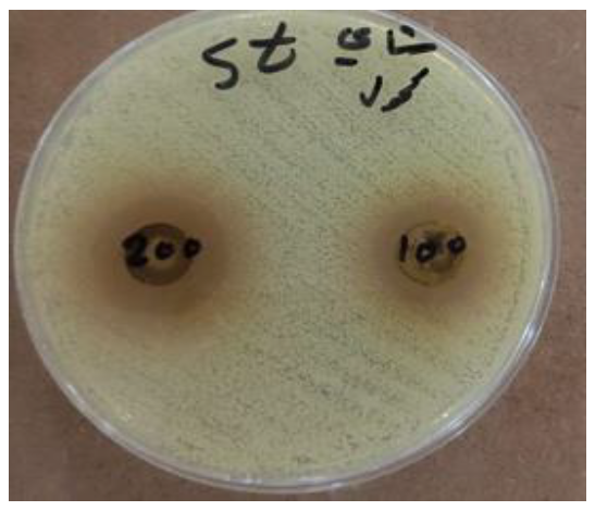
Figure 1: Gel Electrophoresis of Bacteria Isolations
Table 2: Bacterial Isolates and Species Distribution (N = 143 isolates)
Bacterial Species | Number of Isolates | Percentage (%) |
Gram-positive bacteria | 87 | 60.8 |
Staphylococcus aureus | 41 | 28.7 |
Enterococcus faecalis | 15 | 10.5 |
Streptococcus agalactiae | 11 | 7.7 |
Enterococcus faecium | 8 | 5.6 |
Streptococcus pyogenes | 6 | 4.2 |
Staphylococcus epidermidis | 4 | 2.8 |
Streptococcus pneumoniae | 2 | 1.4 |
Gram-negative bacteria | 56 | 39.2 |
Escherichia coli | 22 | 15.4 |
Pseudomonas aeruginosa | 11 | 7.7 |
Klebsiella pneumoniae | 10 | 7.0 |
Enterobacter cloacae | 6 | 4.2 |
Proteus mirabilis | 4 | 2.8 |
Acinetobacter baumannii | 2 | 1.4 |
Citrobacter freundii | 1 | 0.7 |
Infection Pattern | Cases | Percentage (%) |
Monomicrobial infections | 93 | 78.8 |
Polymicrobial infections | 25 | 21.2 |
- Two organisms | 19 | 16.1 |
- Three or more organisms | 6 | 5.1 |
Percentages calculated based on total isolates (n=143) for individual species and total cases (n=118) for infection patterns.
Antimicrobial Susceptibility Patterns
Among S. aureus isolates, 23.4% (10/41) were methicillin-resistant (MRSA) (16,17). All MRSA isolates remained susceptible to vancomycin and linezolid. Clindamycin resistance was observed in 31.7% of S. aureus isolates, with inducible resistance detected in an additional 14.6%. Extended-spectrum β-lactamase (ESBL) production was detected in 31.8% (7/22) of E. coli isolates and 33.3% (1/3) of K. pneumoniae isolates (18,19). Carbapenem resistance was not detected in any Enterobacteriaceae isolates. Fluoroquinolone resistance was present in 27.3% of E. coli isolates [8]. P. aeruginosa isolates demonstrated variable susceptibility patterns, with 18.2% showing multidrug resistance (resistance to three or more antibiotic classes) [9]. All P. aeruginosa isolates remained susceptible to colistin (Table 3).
Table 3: Antimicrobial Susceptibility Patterns of Major Bacterial Isolates
Organism/Antibiotic | Total Tested | Susceptible n (%) | Resistant n (%) |
Staphylococcus aureus (n=41) | |||
Methicillin | 41 | 31 (75.6) | 10 (23.4)* |
Vancomycin | 41 | 41 (100.0) | 0 (0.0) |
Linezolid | 41 | 41 (100.0) | 0 (0.0) |
Clindamycin | 41 | 28 (68.3) | 13 (31.7) |
- Constitutive resistance | 41 | 35 (85.4) | 6 (14.6) |
- Inducible resistance | 41 | 35 (85.4) | 6 (14.6) |
Gentamicin | 41 | 33 (80.5) | 8 (19.5) |
Trimethoprim-sulfamethoxazole | 41 | 37 (90.2) | 4 (9.8) |
Escherichia coli (n=22) | |||
Ampicillin | 22 | 9 (40.9) | 13 (59.1) |
Cefazolin | 22 | 15 (68.2) | 7 (31.8) |
Ceftriaxone | 22 | 15 (68.2) | 7 (31.8)** |
Gentamicin | 22 | 18 (81.8) | 4 (18.2) |
Ciprofloxacin | 22 | 16 (72.7) | 6 (27.3) |
Trimethoprim-sulfamethoxazole | 22 | 17 (77.3) | 5 (22.7) |
Meropenem | 22 | 22 (100.0) | 0 (0.0) |
Klebsiella pneumoniae (n=10) | |||
Ampicillin | 10 | 0 (0.0) | 10 (100.0) |
Cefazolin | 10 | 7 (70.0) | 3 (30.0) |
Ceftriaxone | 10 | 9 (90.0) | 1 (10.0)** |
Gentamicin | 10 | 8 (80.0) | 2 (20.0) |
Ciprofloxacin | 10 | 9 (90.0) | 1 (10.0) |
Meropenem | 10 | 10 (100.0) | 0 (0.0) |
Pseudomonas aeruginosa (n=11) | |||
Ceftazidime | 11 | 8 (72.7) | 3 (27.3) |
Gentamicin | 11 | 9 (81.8) | 2 (18.2) |
Ciprofloxacin | 11 | 8 (72.7) | 3 (27.3) |
Piperacillin-tazobactam | 11 | 9 (81.8) | 2 (18.2) |
Colistin | 11 | 11 (100.0) | 0 (0.0) |
Multidrug resistance*** | 11 | 9 (81.8) | 2 (18.2) |
Molecular Results
The present study demonstrated the presence of specific bacterial DNA bands, as illustrated in the gel electrophoresis image. The bands corresponding to Staphylococcus aureus, Escherichia coli, and Pseudomonas aeruginosa indicate successful amplification of target genes. The migration pattern and intensity of the bands suggest varying levels of DNA concentration among the samples, highlighting the differences in the genetic material present in each bacterial species.
This comprehensive study provides important insights into the bacterial etiology and antimicrobial resistance patterns of circumcision-related infections in pediatric patients. The high rate of bacterial isolation (92.9%) confirms that most circumcision-related infections are indeed bacterial in nature, supporting the routine use of antimicrobial therapy in these cases. The predominance of S. aureus (34.7%) as the leading pathogen is consistent with previous studies and reflects its role as a major skin and soft tissue infection pathogen [8,10]. The presence of MRSA in 23.4% of S. aureus isolates is concerning and highlights the need for routine culture and susceptibility testing rather than relying solely on empirical therapy [11-12]. This finding is particularly relevant given the limited antimicrobial options available in pediatric populations. The significant proportion of Gram-negative bacteria (39.2%), particularly E. coli, suggests possible fecal contamination during or after the procedure [13-14]. This finding emphasizes the importance of proper surgical technique, adequate cleansing of the surgical site, and appropriate post-operative care instructions for caregivers. The detection of ESBL production in nearly one-third of Enterobacteriaceae isolates is alarming and reflects the global trend of increasing antimicrobial resistance [15-16]. These findings have important implications for empirical therapy selection, as first-line β-lactam antibiotics may be ineffective against these organisms [17]. Molecular characterization provided valuable insights into the genetic diversity and virulence potential of isolated pathogens [14]. The presence of biofilm formation genes in a majority of S. aureus and P. aeruginosa isolates may explain treatment failures and delayed healing in some cases, as biofilms can protect bacteria from antimicrobial agents and host immune responses [18-19]. The age-related differences in clinical presentation and bacterial etiology suggest that infection prevention and treatment strategies should be tailored accordingly. Neonates and young infants appear to be at higher risk for S. aureus infections, while older children may be more susceptible to Gram-negative organisms. Several limitations of this study should be acknowledged. The study was conducted in tertiary care hospitals, which may have resulted in selection bias toward more severe cases. In addition, the 18 -month study period cannot capture seasonal variations in bacterial tiology. Exclusion of patients who already receive antimicrobial therapy, although necessary for accurate bacterial isolation, can underestimate by reducing the correct infection rate. The clinical implications of these findings are important. Empirical antimicrobial therapy for circumcised infections should provide coverage for both Gram-positive and gram-negative organisms, assessing local resistance patterns [7,20]. The first generation of cephalosporin may be insufficient as monotherapy, especially in high MRSA or ESBL spread settings. Infection prevention strategies should focus on strict adherence to declining technology, proper surgical storage and strict adherence to extensive instructions after surgery (3.4) during the procedure. Development of standardized protocols for circumcision procedures and post -operative care can help reduce the infection rate. Future research should focus on longitudinal monitoring to monitor bacterial etiology and resistance patterns over time [5-6]. In addition, studies that evaluate the effectiveness of various antimicrobial regimes based on these findings will be valuable for clinical practice.
This study suggests that circumcision infection in children is responsible for a number of bacterial pathogens, with S. Aurous the most prominent organism. Many bacterial species contribute to circumcision infections, with both gram-positive and gram-negative pathogens play an important role. This data strengthens the advice for standard culture -oriented antibiotic therapy in pediatrics associated with circumcision. Empirical therapy should be wide spectrum and modified according to the conclusions of sensitivity. Prevention of these infections and the use of standardized treatment methods can increase the patient's results and reduce the cost of health care related to treatment failure and results.
Morris, B.J. et al. "Estimation of country-specific and global prevalence of male circumcision." Popul Health Metr, 2016, pp. 4.
Weiss, H.A. et al. "Complications of circumcision in male neonates, infants and children: a systematic review." BMC Urol, 2010, pp. 2.
Krill, A.J. et al. "Complications of circumcision." ScientificWorldJournal, 2011, pp. 2458-2468.
Pieretti, R.V. et al. "Late complications of newborn circumcision: a common and avoidable problem." Pediatr Surg Int, vol. 26, no. 5, 2010, pp. 515-518.
World Health Organization. "Antimicrobial resistance: global report on surveillance." Geneva: World Health Organization, 2014.
Friedman, N.D. et al. "The negative impact of antibiotic resistance." Clin Microbiol Infect, vol. 22, no. 5, 2016, pp. 416.
Clinical and Laboratory Standards Institute. "Performance standards for antimicrobial susceptibility testing. 32nd ed. CLSI supplement M100." Wayne, PA: Clinical and Laboratory Standards Institute, 2022.
Tängdén, T. et al. "Foreign travel is a major risk factor for colonization with Escherichia coli producing CTX-M-type extended-spectrum β-lactamases: a prospective study with Swedish volunteers." Antimicrob Agents Chemother, vol. 54, no. 9, 2010, pp. 3564-3568.
Magiorakos, A.P. et al. "Multidrug-resistant, extensively drug-resistant and pandrug-resistant bacteria: an international expert proposal for interim standard definitions for acquired resistance." Clin Microbiol Infect, vol. 18, no. 3, 2012, pp. 268-281.
Bush, K., & Jacoby, G.A. "Updated functional classification of β-lactamases." Antimicrob Agents Chemother, vol. 54, no. 3, 2010, pp. 969-976.
Kaper, J.B. et al. "Pathogenic Escherichia coli." Nat Rev Microbiol, vol. 2, no. 2, 2004, pp. 123-140.
Johnson, J.R., & Russo, T.A. "Extraintestinal pathogenic Escherichia coli: 'the other bad E. coli'." J Lab Clin Med, vol. 139, no. 3, 2002, pp. 155-162.
Otto, M. "Staphylococcus aureus toxins." Curr Opin Microbiol, 2014, pp. 32-37.
Jarraud, S. et al. "Relationships between Staphylococcus aureus genetic background, virulence factors, agr groups, and human disease." Infect Immun, vol. 70, no. 2, 2002, pp. 631-641.
Drews, S.J. et al. "Outbreak of methicillin-resistant Staphylococcus aureus in a neonatal intensive care unit associated with variability in typing results." Am J Infect Control, vol. 33, no. 3, 2005, pp. 139-145.
Bartels, M.D. et al. "A common variant of staphylococcal cassette chromosome mec type IVa in isolates from Copenhagen, Denmark, is not detected by the BD GeneOhm methicillin-resistant Staphylococcus aureus assay." J Clin Microbiol, vol. 47, no. 5, 2009, pp. 1524-1527.
Canton, R., & Coque, T.M. "The CTX-M β-lactamase pandemic." Curr Opin Microbiol, vol. 9, no. 5, 2006, pp. 466-475.
Lowy, F.D. "Staphylococcus aureus infections." N Engl J Med, vol. 339, no. 8, 1998, pp. 520-532.
Costerton, J.W. et al. "Bacterial biofilms: a common cause of persistent infections." Science, vol. 284, no. 5418, 1999, pp. 1318-1322.
Hall-Stoodley, L. et al. "Bacterial biofilms: from the natural environment to infectious diseases." Nat Rev Microbiol, vol. 2, no. 2, 2004, pp. 95-108.
Pitout, J.D., & Laupland, K.B. "Extended-spectrum β-lactamase-producing Enterobacteriaceae: an emerging public-health concern." Lancet Infect Dis, vol. 8, no. 3, 2008, pp. 159-166.
