+91 6002993949
submission@iarconsortium.org
Open Access
ISSN (Print) : 2709-328X
ISSN (Online) : 2709-3298
Sample collection 180 collect samples were collected equally between males and females, from patients attending consulting clinics and suffering from UTI for different ages and both sexes. Urine samples were collected in sterile and sealed plastic tubes and then the tubes were marked with numbers and urine samples were transferred to the laboratory within a period not exceeding 30 minutes. Staphylococcus aureus is one of the most important and most common pathogenic species for humans. Species belonging to the genus are responsible for a wide range of diseases such as boils, impetigo and wound abscesses resulting from surgery. Pathogenicity and its ability to invade the host's tissue and spread in it is due to its possession of many virulence factors. Genetic elements called plasmids are in the form of circular ring structures of a double strand of DNA. They have the ability to be inherited steadily and spread widely in the cells of different bacteria. PCR technique was used to investigate the hemolysin gens possessed by bacterial isolates. The HLA gene is the main virulence factor encoding its pathogenicity. It is active against a group of host cells, including red blood cells. It gives an indication of the role of these genes in the pathogenesis of Staphylococcus aureus among hospitalized patients. Hemolysin is one of the most important virulent agents for bacteria that cause infections outside the intestine. The hemolysin enzyme breaks down the membranes of the red blood cells by making small holes in their membranes. It provides the bacteria with the iron they need, and as a result of its toxicity to the cells, leads to the destruction of the host's kidney tissues.
The urinary system is the one responsible for storing and disposing of urine in addition to its disposal of waste products excreted by the kidneys The kidneys form urine by filtering dirt and excess water from the blood.
Then its journey begins from the kidneys through two thin tubes called Ureters to reach the bladder, and when the bladder is full, a person must urinate through the urethra and for the urinary system to function, it needs to cooperate with the lungs, skin and intestines, in order to maintain the chemical and water balance in the body. Adults get rid of between 800 and 2,000 ml of daily fluids when consuming the recommended daily amount of fluids of up to 2 liters, according to the National Institutes of Health But in some cases, in which certain types of drugs, such as those used to treat high blood pressure, are consumed, the amount of urine that this person produces is affected accordingly, and some types of drinks, that is, those that contain caffeine generate more urine because they are generating it.
Urinary tract infections are one of the health problems that afflict a large proportion of the human community, estimated in the millions annually [1].
Urinary tract infections are also the most common cause of hospital infection, accounting for up to 35% of hospital-acquired infections.
Females are more susceptible to infection than males because of their short urethra compared to males and its proximity to the hot and humid area surrounding the rectum that is teeming with microorganisms, where bacteria can reach the bladder more easily in females [2]. A urinary tract infection affects any part of the urinary system; That is, the kidneys, ureter, bladder, and urethra, and women are often more susceptible to urinary tract infection than men, and infection limited to the bladder is painful and annoying, and severe consequences can occur if it moves to the kidneys and urinary tract infection occurs more often at a rate of 8: 1 among men and women This means that for every 8 women with a UTI, there is only one man who has the disease.
Urinary Tract Infection Risk Classifications
Upper Organ Infection: in the kidneys
Lower System Infection: that is, in the urinary
bladder, urethra and prostate
There is a clear distinction between simple urinary tract infection, that is, an infection in the lower urinary tract in a non-pregnant woman, and complex infection (all the rest). Every infection in a man, as well as an infection in the kidneys, is a complex infection. An infection of this type requires certain tests and specialist intervention [3].
The Most Common Infection
Of all human infections, UTIs are the most common and require antibiotic treatment. The percentage of infections increases with age, and it is more prevalent among women: at the age of 16-35 it reaches 20%, and at the age of 65 and over it reaches 40%. The rate of infections in men, up to the age of 35, is less than 1% and increases to 20% and more over the age of 65, as a result of an enlarged prostate.
Urinary tract infection is usually an ascending infection, meaning that the bacteria (bacteria) causing the infection originate in the woman's vulva (vulva) and in the anus (anus). The occurrence of infection depends on the virulence and type of germ, as well as in the factors related to the patient. For example, complete bladder emptying is of great importance. Due to factors such as an enlarged prostate in a man, or a prolapsed uterine (uterine) in a woman, the emptying is not complete. The infection may stem from a defect in the valve between the ureters and the urinary bladder, from the presence of stones in the urinary system, or from every congenital defect in the urinary system.
Role and Structure of Hemolysin
Antibodies to surface erythrocyte antigens, able to destroy erythrocyte membranes with the participation of a supplement, as a result of which hemoglobin is released into the surrounding solution - hemolysis. They appear in serum during immunization with foreign erythrocytes and autoimmune diseases. G. - The cause of intravascular hemolysis during incompatible and hemolytic transfusion. Newborn diseases. G. It is called. Also toxins of microorganisms (staphylococcus, streptococcus, etc.), which cause enzymatic destruction of red blood cells [4].
Hemolysin is an enzyme that affects cells and is characterized by its ability to analyze red blood cells and play a role in urinary tract infections through its toxicity to the epithelial cells of the kidney .
Hemolysin is classified into three types depending on its ability to analyze red blood cells and pathological events, the first type analyzes the membranes and is called the beta-hemolysis, while the second type is holes in the cell membrane, and a green area appears around the colony and it is called Alpha-hemolysis, while the third type breaks down the cell wall and is called gamma-hemolysis, and there are some types that do not show decomposition around colonies and are called non-hemolytic bacteria .
Hemolysin is a type of cytolytic enzyme, as it works to break down urinary epithelium cells. After attaching to these cell membranes, it creates holes in them that cause ATP to exit from them, and then cell death.
The mechanisms that produce hemolysin, which cause infection and stimulate the kidney (Hemolysin enzyme is a pore-forming cell lysate and acts as an important virulence agent in intestinal and extra-intestinal Escherichia coli Or it is a toxic protein that lyses cells by forming holes in the target cell wall and affects red blood cells, white blood cells, and renal tubular cells This enzyme is an important virulent factor in causing bacterial infections. It is produced by some types of bacteria to analyze red blood cells, and it has a different molecular structure according to the type of bacteria, An enzyme secreted outside the bacterial cell during the logarithmic phase and in which its degradation is partial, and the second type beta-hemolysin is an enzyme associated with the cell and its degradation is complete, and the efficacy of this type cannot be separated from the metabolic activities of the cell The third type is Kama. - Hemolysin, as this enzyme breaks down the cell wall, and there are other types of bacteria that do not show degradation around the colony, and these types are called non-hemolytic bacteria
Coli bacteria also produce the enzyme enter hemolysin, which is a cell toxin produced by bacteria during the logarithmic phase of growth and accumulates inside the cell The enzyme alpha-hemolysin is the most common type produced by the E. coli bacteria, and it is not bound to the cell.
The process of secreting the hemolysin enzyme by E. coli bacteria takes place in two ways, the first by gathering the hemolysin enzyme in the bacterial cytoplasmic membrane, and this process requires the energy of the hlyB genes, and the second releases the hemolysin enzyme from the outer membrane to the periphery, and this process requires heat and the hlyD gene and does not require energy.
Dry Hot Sterilization
All glassware used in an electric oven (Oven) were sterilized at 168 ° C for an hour and a half
Wet Hot Sterilization
The ready-made and synthetic agricultural media that are not affected by heat were sterilized with Autoclave at 121 ° C at one pressure of atmosphere for 15 minutes C
Filtration Sterilization
Heat-sensitive materials and solutions were sterilized using millipore_filters with a diameter of 0.22 μm.Preparation of solutions and reagents
Preparation of Solutions and Reagents Solutions Normal Saline: By dissolving 0.85 grams of NaCl in 90 ml of distilled water Complete the volume to 100 ml, then sterilize it with the oxidizer
BaCL2
McFarland Standard Tube (0.5). Mcfarland Tube Standard No. Prepared from the following solutions as stated in Benson (2002): A - Barium chloride dilute tube 1.175 grams of barium chloride in 90 mL of sterile distilled water, complete the volume to 100 ml for 0.048 concentration H2SO4 - Sulfuric acid solution (Figure 1-2).
H2SO4 Add 1 ml of H2SO4 sulfuric acid to 90 ml sterile distilled water, and complete the volume to 100 ml. When using a McFarland prolonged, mix 0.5 mL of barium chloride solution with 99.5 mL of sulfuric acid solution to obtain an approximate number of bacterial cells of 1.5 *

Figure 1: Laboratory Centrifuge/Shaker for Sample Processing

Figure 2: Vortex Mixer for Efficient Sample Mixing in Laboratories
10 milliliter cells. Distribute the solution to sterile test tubes with 4 mL seals per tube and store in dark places At room temperature, the tube was used for the purpose of comparing the density of the bacterial growth in the inoculum used with the density The solution is in the tube.
Phosphate Buffer Solution
The solution (PBS) was prepared by dissolving 18 mg of sodium chloride, 0.34 mg of potassium dihydrogen phosphate and 1.12 mg of potassium dihydrogen phosphate in 100 ml of distilled water and adjusting the pH to 7.3. Sterilize the solution with oxide.
Bacterial Genomic DNA
The bacterial nucleic acid was extracted by using the Genomic DNA Extraction kit provided by the American company Geneaid, and the extraction was carried out according to the manufacturer's instructions:
Transfer 1 ml of suspensions from each growing bacterial isolate to the medium of broth, infuse the heart and brain, and put sterile 1.5 ml tubes of Abendorff, then transfer them to a centrifuge and discard at a speed of 15,000 minutes for one minute in order to collect the bacterial cells and then discard the floating liquid
Add 200 microliters of Lysozyme Buffer (20mg / ml) and mix the mixture with a vortex mixer for 5 seconds
Incubate the mixture at room temperature for 10 minutes, and during the incubation period, invert the tubes to ensure complete lysis of cells in the mixture
Genetically speaking, the PCR technique was used to investigate the hemolysin gens possessed by bacterial isolates, as DNA was extracted using the kit used for this purpose and electrophoresis on the acerose gel (1.5%) and was detected by using the ethidium bromide dye and examined under ultraviolet rays (UV).). For staph bacteria. aureus, 10 isolates were selected to investigate the hemolytic genes, and the primers amplification products showed that all tested isolates contained 100% of the gene encoding the production of hemolysin with a product of size 190 bp, and 40% of the bacterial isolates contained the gene with a size of 293 bp, and this It gives an indication of the role of these genes in the pathogenesis of Staphylococcus aureus among hospitalized patients. The HLA gene is the main virulence factor encoding its pathogenicity (Figure 5-7). It is active against a group of host cells, including red blood cells (Figure 3-4 and Table 1-2).
Table 1: The Number of Samples for Bacterial Presence
| Result | Number of samples | Percentage |
| 66.68 | 122 | Positive sample |
| 33.32 | 58 | Negative sample |
| 100 | 180 | Total |
Table 2: The Number of Bacteria Distributed According To the Analysis
| Bacterial isolates | N |
| Hemolytic | 50 |
| Non-hemolytic | 70 |

Figure 3: Explain the Percentage of Number of Samples
Figure 4: Bacteria Isolated From Urinary Tract Infections
Figure 5: Percentage of Bacteria Distributed According To the Analysis
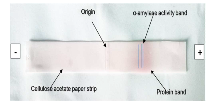
Figure 6: Bacterial Isolates According To Their Ability to Produce Hemolysin
Figure 7: Resistant Isolates Bacteria of Antibiotics
It works Hemolysin is one of the most important virulent agents for bacteria that cause infections outside the intestine. It is a different type of cell, such as granular white blood cells, lymphocytes, red blood cells, and renal tubular cells [5]. The hemolysin enzyme breaks down the membranes of the red blood cells by making small holes in their membranes, as the presence of hemolysin is an important factor in providing the bacteria with the iron they need, and as a result of its toxicity to the cells, it leads to the destruction of the host's kidney tissues, as the hemolysin produced from some strains of E. coli is an important virulence factor in many infections, such as pelvic and renal infection and septicemia. May and his group (2000) indicated that strains incapable of producing hemolysin are less virulent and pathogenic than those that produce it As most strains of this bacterium present in a natural flora in the human intestine do not have the ability to produce hemolysin, but once it colonizes the urinary tract, it becomes a producer of it.
The results of the anti-biotic susceptibility test for the mutant cells also indicated the transfer of the resistance to antibiotics and the transformation of the mutated cells from susceptible to resistant to the antibiotics used in the study if the results showed the transmission of the anti-resistance trait of ampicillin, methicillin.
Tobramycin and bacitracin for coli cells. E mutant. Several studies have proven that the resistance of S. aureus bacteria for most of the antibiotics encodes for it genes carried on the plasmid or transposons, in addition to the ability of some types of plasmids to transfer from one strain to another naturally through the process of bacterial conjugation, carrying with it the characteristic of antibody resistance. And this is what may explain The prevalence of antibiotic resistance among the genera of bacteria (the degradation test of casein showed the ability of the aureus S bacteria to analyze casein through the appearance of the transparent halo around the hole due to its ability of the bacteria to produce T proteins with L to analyze casein similar in its composition to fibrin - in the presence of blood plasma) One of these proteins which is secreted by aureus. It is used as a solvent for blood clots, and that the absence of the transparent aura of the mutant cells may give an indication that the gene encoding this protein is carried on the chromosome of the bacterial cell and not on the plasmids.
Kong, C. et al. “Targeting Staphylococcus aureus Toxins: A Potential Form of Anti-Virulence Therapy.” Toxins, vol. 8, no. 3, 2016, p. 72.
Monecke, S. et al. “Staphylococcus aureus In Vitro Secretion of Alpha Toxin (hla) Correlates with the Affiliation to Clonal Complexes.” PLOS One, vol. 9, no. 6, 2014, e100427.
O’Callaghan, R.J. et al. “Specific Roles of Alpha-Toxin and Beta-Toxin during Staphylococcus aureus Corneal Infection.” Infection and Immunity, vol. 65, no. 5, 1997, pp. 1571–1578.
Osman, K. et al. “Antimicrobial Resistance and Virulence Characterization of Staphylococcus aureus and Coagulase-Negative Staphylococci from Imported Beef Meat.” Annals of Clinical Microbiology and Antimicrobials, vol. 16, no. 1, 2017, pp. 1–10.
Xiao, M. et al. “Genotypic Diversity of Staphylococcus aureus α-Hemolysin Gene (hla) and Its Association with Clonal Background: Implications for Vaccine Development.” PLOS One, vol. 11, no. 2, 2016, e0149112.
