+91 6002993949
submission@iarconsortium.org
Open Access
ISSN (Print) : 2709-328X
ISSN (Online) : 2709-3298
Liposuction is a well-known cosmetic surgical technique performed by plastic surgeons to achieve ideal body contouring by removing excess adipose tissue. Despite being considered a safe procedure, liposuction may result in complications, particularly in patients with comorbidities such as cardiovascular or pulmonary diseases, a history of smoking, or when combined with other surgical procedures. We present a case of a 36-year-old female (BMI 26 kg/m²) who underwent Power-Assisted Liposuction (PAL) (MicroAire) without the use of an energy-based device, followed by a tummy tuck. Postoperatively, the patient developed skin necrosis, which was managed conservatively with Lovenox, calcium channel blocker cream, and later mechanical dermabrasion with tranexamic acid injection. The necrotic area resolved completely within several weeks, and the patient recovered well. This case raises awareness that PAL (MicroAire) can be aggressive and may lead to necrosis, contrary to manufacturer claims of safety.
Liposuction is the second most commonly performed cosmetic procedure in the United States. Its primary goal is to enhance body contour by removing excess subcutaneous fat. This procedure plays a vital role in limiting future fat deposition in targeted areas and reducing Body Mass Index (BMI). Additionally, liposuction allows plastic surgeons to redistribute volume according to patient preferences, offering lower complication rates than other surgical interventions. Although complications are relatively rare, certain risk factors increase the likelihood of postoperative issues, including cardiovascular disease, pulmonary disease, vascular conditions, and tobacco use. Common complications include seroma, hematoma, edema, ecchymosis, venous thromboembolism, and, rarely, skin necrosis. We present a case of a 36-year-old female (BMI 26 kg/m²) who underwent PAL (MicroAire) followed by a tummy tuck and developed postoperative skin necrosis at the left flank and tummy tuck scar. Conservative treatment led to full recovery, demonstrating that PAL alone can potentially cause necrosis, even in the absence of an energy-based device.
Case Presentation
A 36-year-old healthy female with a past surgical history of two rhinoplasties and a BMI of 26 kg/m² presented for PAL (MicroAire) and tummy tuck. The patient was a non-smoker with no allergies. Preoperatively, anatomical landmarks were marked, and routine antisepsis with chlorhexidine was performed. The surgical field was draped in a sterile manner. A tumescent solution containing adrenaline (2 mg/L) and lactated Ringer’s solution was infiltrated into the subcutaneous adipose tissue. Following induction of anesthesia, a 2-3 mm stab incision was made to allow access for the liposuction cannula (30 cm in length, 5 mm in diameter, Mercedes tip). The PAL cannula was connected to a motorized handpiece and inserted in a deep-to-superficial plane using a systematic, multidirectional fanning technique. Suction was applied via a high-powered vacuum pump, ensuring uniform fat removal and smooth contouring. After PAL, a tummy tuck was performed, leaving a small 2 cm vertical scar below the new umbilicus. On postoperative days 1 and 2, skin necrosis became evident on the tummy tuck scar and left flank (Figure 1).
Treatment included Lovenox (low molecular weight heparin) injections around the affected area to improve blood flow and calcium channel blocker cream application. The patient was discharged and followed up regularly. After one-month, mechanical dermabrasion was performed with a sterile Pierre Ponce, leading to significant improvement (Figures 2-5).
Figure 1: Skin necrosis post-surgery
Figure 2: Skin necrosis at the tummy tuck scar in stages of improvement

Figure 3: Skin necrosis on the left flank in stages of improvement

Figure 4: Significant improvement of skin necrosis on the flank after dermabrasion

Figure 5: Significant improvement of skin necrosis below the tummy tuck scar after dermabrasion
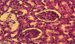
Figure 6: Complete resolution of the necrotic area on the left flank several weeks post-surgery
Figure 7: Complete resorption of the necrotic area at the tummy tuck scar several weeks post-surgery
One week after dermabrasion, an injection of tranexamic acid was administered to enhance wound healing. Complete resolution of the necrotic areas was observed within several weeks (Figures 6-7).
Liposuction has become one of the most widely performed cos metic procedures worldwide. The primary objective of this surgical technique is to reduce fat deposits in targeted areas of the body, enhancing overall body contouring with minimal recovery time. This procedure involves the insertion of an aspiration cannula through small skin incisions, utilizing suction to remove fat deposits efficiently. Although generally regarded as a safe surgical intervention, liposuction is not devoid of risks, as it can lead to various post-operative complications. These complications are broadly categorized into systemic and local effects. Systemic complications encompass conditions such as hypothermia, significant blood loss, visceral perforation, and severe infections, including necrotizing fasciitis and toxic shock syndrome. Additionally, fat embolism, thromboembolism, deep vein thrombosis, and pulmonary edema are recognized as potential life-threatening risks associated with liposuction. On the other hand, local complications include predictable post-operative effects such as edema and ecchymosis. Other possible local adverse effects involve the formation of seromas and hematomas, wound infections, and contour irregularities, such as over-correction-here excessive fat removal surpasses the desired contour line and under-correction, leaving residual fat deposits in the treated areas. Moreover, asymmetry, skin laxity, hyperpigmentation, umbilical deviation, and sensory disturbances like hypoesthesia, which frequently occur following liposuction, may also be observed. Although rare, more severe complications such as neuroma formation and skin necrosis can occur [1-3]. Fayi et al. [4] documented the case of a 31-year-old Saudi female who presented with unilateral right eye vision loss, bruises, and burning pain in areas where she had undergone liposuction. Notably, the patient was a smoker and was taking oral contraceptives, which likely contributed to her development of retinal vein thrombosis. Initially managed conservatively, she was discharged but returned five days later with infected necrotic surgical sites. The severity of her condition necessitated multiple debridements and grafting to manage the affected areas [4]. Another case, reported by Tan et al. [5], described a 20-year-old female who underwent bilateral thigh liposuction. The patient had no prior medical history, was a non-smoker, and was not obese. However, on the seventh post-operative day, she developed signs of cutaneous fat necrosis, particularly in the upper thigh region, which became evident upon removing her dressing and elastic garment. Within two weeks, the necrotic process progressed, leading to multiple full-thickness skin necrosis lesions without any apparent signs of infection. The management plan included conservative treatment with regular dressings, and following the removal of residual necrotic fat and thorough irrigation, the wounds were closed by secondary suturing. Ultimately, the skin necrosis resolved completely within 60 days [5]. Similarly, Nseir et al. [6] reported a case involving a 42-year-old woman with no prior medical conditions who underwent thigh liposuction performed by a dermatologist. Post-operatively, she developed skin necrosis on the lateral thigh, which necessitated surgical necrosectomy in the operating room. Fortunately, her wound healed completely within two weeks following surgical intervention [6]. A study conducted by Yang et al. [7] evaluated the efficacy and complications of four modified Liposuction-Curettage (LC) methods for treating Axillary Bromhidrosis (AB) in a cohort of 280 patients, 228 of whom completed follow-up. The surgical approach included mini-incisions, tumescent LC, and meticulous post-operative care. The results demonstrated that well-executed LC techniques, combined with post-surgical management strategies such as skin pinching and appropriate dressings (cotton balls, gauze, and elastic bandages), effectively eliminated malodor in 89.81% of cases. Notably, minor complications included small skin necrosis (2.55%) and epidermal damage (17.83%) [7]. Zhitny et al. [8] detailed the case of a 47-year-old female who experienced abdominal scar dehiscence following a failed tummy tuck procedure performed in Mexico. Upon evaluation, the wound was debrided, and necrotic tissue was excised. One month later, a second debridement and wound closure were performed successfully, leading to complete recovery and proper wound healing by post-operative day 30, without any further complications [8]. A similar case of skin necrosis following abdominoplasty was also documented by Zhitny et al. [9]. To our knowledge, our case represents the first reported instance of Power-Assisted Liposuction (PAL) using the MicroAire device complicated by acute skin necrosis, which remains an exceedingly rare adverse event associated with this surgical procedure. Despite the manufacturer’s claims that PAL is safe and poses no risk of necrosis, our case contradicts this assertion, as necrosis developed despite the absence of energy-based devices such as Vibration Amplification of Sound Energy at Resonance (VASER) liposuction. Furthermore, while most cases of post-liposuction necrosis require adjunctive therapies, including hyperbaric oxygen sessions, carboxytherapy, and wound Vacuum-Assisted Closure (VAC) to promote tissue healing, these modalities were not necessary in our case.
Instead, conservative management with Lovenox injections and calcium channel blocker cream resulted in successful resolution, with complete healing achieved without the need for surgical debridement.
Liposuction remains an essential tool in body contouring, but it carries risks that must not be overlooked. Our case highlights the rare but serious complication of skin necrosis following PAL (MicroAire) in a non-smoker, healthy patient with no predisposing risk factors. Despite PAL being marketed as a safer alternative to energy-based liposuction techniques, our findings indicate that it can still lead to necrosis, particularly when combined with a tummy tuck. Fortunately, conservative management resulted in complete wound healing without surgical debridement. Further research and case studies are necessary to better understand the risk factors and optimize treatment strategies for post-liposuction skin necrosis.
Wu, Shannon, et al. “Liposuction: Concepts, safety, and techniques in body-contouring surgery.” Cleveland Clinic Journal of Medicine, vol. 87, no. 6, June 2020, pp. 367-375. http://dx.doi.org/10.3949/ccjm.87a.19097.
Bustos, Raul Cuevas, et al. “Necrotizing soft tissue infection after liposculpture; case report.” International Journal of Surgery Case Reports, vol. 77, 2020, pp. 677-681. http://dx.doi.org/10.1016/j.ijscr.2020.11.078.
Skorochod, Ron, et al. “Perforation of abdominal viscera following liposuction: A systemic literature review.” Aesthetic Plastic Surgery, vol. 46, no. 2, August 2022, pp. 774-785. http://dx.doi.org/10.1007/s00266-021-02532-9.
Fayi, Khalid A, et al. “The incident of multiple skin necrosis and unilateral vision loss post liposuction: A case report.” Cureus, vol. 15, no. 6, June 2023. http://dx.doi.org/10.7759/cureus.40384.
Tan, Liuchang and Yuangang Lu “Severe thigh skin necrosis after liposuction.” Clinical & Experimental Dermatology and Therapies, vol. 9, no. 1, February 2024. http://dx.doi.org/10.29011/2575-8268.100219.
Nseir, Iad, et al. “Skin necrosis of the thigh after a cryolipolysis session: A case report.” Aesthetic Surgery Journal, vol. 38, no. 4, February 2018, pp. NP73-NP75. http://dx.doi.org/10.1093/asj/sjx028.
Yang, Hu, et al. “Effectiveness and complications of improved liposuction–curettage through mini-incisions for the treatment of axillary osmidrosis.” Plastic Surgery, vol. 25, no. 4, September 2017, pp. 234-241. http://dx.doi.org/10.1177/2292550317728038.
Zhitny, Vladislav Pavlovich, et al. “Abdominal flap necrosis and wound dehiscence following a medical tourist tummy tuck.” Case Reports in Surgery, vol. 2020, November 2020, pp. 1-4. http:// dx. doi. org/ 10.1155/ 2020/ 8819102.
Zhitny, Vladislav Pavlovich, et al. “Anatomic reconstruction for major tissue loss following abdominoplasty: A case report.” International Journal of Surgery Case Reports, vol. 72, June 2020, pp. 241-244. http://dx.doi.org/10.1016/j.ijscr.2020.06.019.
