We present a rare case of an 11-year-old girl with an unusual clinical presentation of bluish patches on the trunk and legs accompanied by gum bleeds, ultimately diagnosed as TB-associated secondary ITP. Despite the absence of typical TB symptoms and negative serological tests, a lymph node biopsy revealing necrotic lymph nodes positive for AFB, along with a reactive tuberculosis skin test, confirmed the TB diagnosis, leading to anti-tubercular therapy initiation. This case emphasizes the importance of considering TB as an underlying etiology in secondary ITP, particularly in regions with high TB prevalence, highlighting diagnostic challenges and the need for early intervention to prevent complications.
Keywords
Tubercular lymphadenitis
Immune Thrombocytopenic Purpura (ITP)
INTRODUCTION
Immune thrombocytopenic purpura (ITP), characterized by a decrease in platelet count and mucocutaneous bleeding, is a hematologic disorder frequently encountered in pediatric practice. ITP can either be primary (idiopathic) or secondary, with the latter often attributed to various underlying causes. One rare and less explored etiological association is the coexistence of ITP with tuberculosis (TB) [1,2].
Tuberculosis, a major global health concern, typically affects the lungs but can manifest in extrapulmonary sites, posing diagnostic challenges. Tubercular lymphadenitis, characterized by lymph node enlargement, is one such extrapulmonary manifestation of TB that may go unnoticed or misdiagnosed due to its varied clinical presentations. Here, we present a remarkable case of an 11-year-old adolescent girl with a unique presentation of tubercular lymphadenitis coexisting with secondary ITP [3,4].
In pediatric patients, the coexistence of TB and ITP is an infrequent clinical scenario, and to the best of our knowledge, this case represents a rare occurrence with limited documentation in the medical literature. Understanding the underlying pathophysiological mechanisms linking TB and ITP, along with their diagnostic challenges and clinical management, is crucial in effectively managing such complex cases [5,6].
This case report aims to shed light on the intriguing intersection of tubercular lymphadenitis and secondary ITP, emphasizing the importance of considering TB in the differential diagnosis of secondary ITP, particularly in regions with a high TB burden. Moreover, it underscores the significance of prompt diagnosis and timely initiation of appropriate treatment for both conditions, thus preventing unnecessary morbidity and complications.
In this context, we discuss the clinical presentation, diagnostic workup, and management of this unique case, which presented with bluish patches, gum bleeds, and cervical lymphadenopathy, ultimately leading to the diagnosis of TB-associated secondary ITP. Through this case, we highlight the importance of considering TB as an underlying etiology in secondary ITP and the challenges faced in diagnosis and management, particularly in pediatric patients.
In the era of increasing multidrug-resistant TB and the ongoing global efforts to control this infectious disease, recognizing and managing cases of TB-associated secondary ITP are of paramount importance. This report serves as a valuable addition to the existing literature, offering insights into the intricate interplay between TB and ITP and providing guidance for clinicians faced with similar diagnostic conundrums.
Case Report
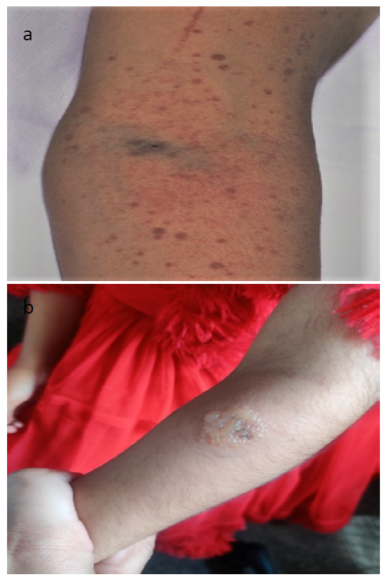
We present the case of an 11-year-old adolescent girl who came to our attention with complaints of bluish patches appearing on her trunk and legs for the past 15 days, accompanied by gum bleeds persisting for the last 3 days. The patient denied experiencing gross hemoptysis, hematemesis, epistaxis, melena, or hematuria. There was no past medical history suggestive of hematological or liver diseases, and the patient had not been on recent medications. Notably, there was a significant history of contact with her grandmother, who had been undergoing treatment for sputum-positive tuberculosis for the preceding 6 months. Physical examination revealed widespread purpura with pronounced oozing of blood from the gums. Additionally, we noted a diffuse swelling in the left side of her neck, comprising several mobile, non-tender, and firm lymph nodes, measuring between 1 to 3 cm in diameter. Examination of the spleen and liver did not yield any palpable abnormalities, and the remainder of the physical examination was unremarkable (Figure 1).
Figure 1: a) Clinical Presentation of Bluish Patches, b) Tuberculosis Skin Test Positive
Diagnostic Evaluation
In pursuit of the underlying etiology, we conducted a battery of serological tests, including hepatitis A, B, C, HIV, and Epstein–Barr virus, along with an autoimmune profile encompassing antinuclear antibody, complement C3, and C4 levels. The results of all these tests returned negative. Subsequently, a lymph node biopsy was performed, which revealed necrotic lymph nodes and tested positive for acid-fast bacilli (AFB). Contrary to our expectations, the Cartridge-Based Nucleic Acid Amplification Test (CBNAAT) did not support a diagnosis of tuberculosis. However, a tuberculosis skin test was conducted, yielding a reactive result with an 18 mm induration. Based on these findings, the patient was promptly initiated on anti-tubercular therapy.
DISCUSSION
Immune thrombocytopenic purpura (ITP) is a well-recognized hematologic disorder characterized by a decreased platelet count and mucocutaneous bleeding. In pediatric practice, primary ITP is more common, often occurring after viral infections. Secondary ITP, on the other hand, is associated with underlying conditions, including autoimmune diseases, infections, and malignancies. Tuberculosis (TB) is a rare but potentially overlooked etiological association with secondary ITP, and it poses unique diagnostic and management challenges [2-4].
Tuberculosis is a global health concern with diverse clinical manifestations. Pulmonary TB is the most common form, but extrapulmonary manifestations can be elusive and may result in diagnostic delays. Tubercular lymphadenitis, characterized by the enlargement of lymph nodes, is one such extrapulmonary manifestation that can mimic other conditions, making it challenging to diagnose. In our case, the adolescent girl presented with generalized purpura, gum bleeds, and cervical lymphadenopathy, creating a diagnostic puzzle [5-7].
The coexistence of TB and ITP, particularly in pediatric patients, is a rarity with limited documentation in medical literature. This case highlights the importance of considering TB in the differential diagnosis of secondary ITP, especially in regions with a high TB burden. The mechanism underlying the association between TB and ITP remains unclear but may involve immune dysregulation triggered by TB infection [6-8].
Diagnosing TB-associated secondary ITP can be challenging due to the diverse clinical presentation of TB. In our case, serological tests and autoimmune profiles returned negative results, illustrating that TB-associated ITP may not be accompanied by typical laboratory abnormalities seen in primary ITP or other secondary ITP etiologies. A lymph node biopsy provided crucial diagnostic information by revealing necrotic lymph nodes and AFB positivity. However, it is noteworthy that the Cartridge-Based Nucleic Acid Amplification Test (CBNAAT) did not support TB diagnosis, highlighting the complexity of TB diagnostics. The positive tuberculosis skin test with an 18 mm induration further supported the TB diagnosis. This emphasizes the importance of considering a combination of clinical, radiological, and laboratory findings when assessing TB in secondary ITP cases [9-10].
Timely diagnosis and management of both TB and ITP are crucial in preventing complications. In our case, the patient was promptly initiated on anti-tubercular therapy, targeting the TB component of her condition. Managing the ITP component involves addressing the thrombocytopenia and associated bleeding symptoms. Corticosteroids, intravenous immunoglobulins (IVIG), and platelet transfusions are therapeutic options to consider. Close monitoring of platelet counts and bleeding symptoms is essential to adjust treatment as needed.
CONCLUSION
This case underscores the importance of recognizing the intersection of TB and ITP in pediatric patients. It highlights the complexity of diagnosis and the necessity of considering TB as an underlying etiology in secondary ITP, especially in regions with a high TB burden. Multidisciplinary collaboration between pediatricians, hematologists, and infectious disease specialists is essential to ensure timely and appropriate management of both conditions. Moreover, this case serves as a valuable contribution to the medical literature, raising awareness of this unique clinical scenario and providing guidance for clinicians facing similar diagnostic challenges in TB-endemic areas.
REFERENCE
Kühne, T. et al. “A prospective comparative study of 2540 infants and children with newly diagnosed idiopathic thrombocytopenic purpura from the Intercontinental Childhood ITP Study Group.” Journal of Pediatrics, vol. 143, no. 5, 2003, pp. 605–608.
Dhingra, K.K. et al. “Tuberculosis masquerading as chronic idiopathic thrombocytopenic purpura: Case report and review of literature.” Indian Journal of Hematology and Blood Transfusion, vol. 30, 2014, pp. 105–108.
Javed, S. et al. “Immune thrombocytopenic purpura as a presenting feature of tuberculosis.” Journal of the College of Physicians and Surgeons Pakistan, vol. 19, no. 4, 2009, pp. 244–245.
Garcia, D. and Erkan, D. “Diagnosis and management of the antiphospholipid syndrome.” The New England Journal of Medicine, vol. 378, no. 21, 2018, pp. 2010–2021.
Eshghi, P. et al. “Prevalence of hepatitis C virus genotypes in Iranian patients.” Archives of Iranian Medicine, vol. 10, no. 3, 2007, pp. 352–355.
Jain, R. et al. “Tubercular lymphadenitis presenting as immune thrombocytopenic purpura.” Indian Journal of Pediatrics, vol. 80, no. 2, 2013, pp. 175–177.
Choudhury, S.R. et al. “Unusual presentation of childhood tuberculosis.” Indian Journal of Pediatrics, vol. 78, no. 11, 2011, pp. 1414–1415.
Pandey, S. et al. “Disseminated tuberculosis presenting as immune thrombocytopenic purpura in a child.” Journal of Tropical Pediatrics, vol. 59, no. 3, 2013, pp. 234–236.
Sharma, S.K., Mohan, A. and Sharma, A. “Mitochondrial dysfunction in immune thrombocytopenic purpura: Implications for pathogenesis and treatment.” European Journal of Haematology, vol. 80, no. 5, 2008, pp. 488–491.
Dhanrajani, A. et al. “Isolated thrombocytopenia: An unusual presentation of tuberculosis.” Journal of Pediatric Hematology/Oncology, vol. 25, no. 2, 2003, pp. 160–163.
Recommended Articles
Case Report
OROFACIAL GRANULOMATOSIS: A CASE REPORT
Diksha Thakur,
...
Mandeep Kaur
Published: 19/11/2024
Download PDF
Research Article
The Study of Suicide in Adolescents in The Context of Cognitive Brain Change
Zhang Wen,
...
Amiya Bhaumik
Published: 29/07/2024
Download PDF
Research Article
Role of First Trimester HbA1c in Screening of Gestational Diabetes Mellitus
Anu Berwal,
...
Geetika Gupta Syal
Published: 28/12/2025
Download PDF
Case Report
The Utility of Maternal Serum Amyloid a in the Prediction of Preterm Birth in Symptomatic Women
Enas Hussein Ahmed,
Aseel Ghazi Rifat
Published: 30/06/2024
Download PDF
PO Box 101, Nakuru
Kenya.
Email: office@iarconsortium.org
Editorial Office:
J.L Bhavan, Near Radison Blu Hotel,
Jalukbari, Guwahati-India
None, C. & Kapoor, P. (2023). Unveiling the Puzzle: Tuberculosis-Associated Secondary Immune Thrombocytopenic Purpura in an Adolescent with Unique Clinical Presentations. Scientific Research Journal of Medical Case Reports, 3(2), 1-3.
MLA
None, Chetna and Pankaj Kapoor. "Unveiling the Puzzle: Tuberculosis-Associated Secondary Immune Thrombocytopenic Purpura in an Adolescent with Unique Clinical Presentations." Scientific Research Journal of Medical Case Reports 3.2 (2023): 1-3.
Chicago
None, Chetna and Pankaj Kapoor. "Unveiling the Puzzle: Tuberculosis-Associated Secondary Immune Thrombocytopenic Purpura in an Adolescent with Unique Clinical Presentations." Scientific Research Journal of Medical Case Reports 3, no. 2 (2023): 1-3.
Harvard
None, C. and Kapoor, P. (2023) 'Unveiling the Puzzle: Tuberculosis-Associated Secondary Immune Thrombocytopenic Purpura in an Adolescent with Unique Clinical Presentations' Scientific Research Journal of Medical Case Reports 3(2), pp. 1-3.
Vancouver
Chetna C, Kapoor P. Unveiling the Puzzle: Tuberculosis-Associated Secondary Immune Thrombocytopenic Purpura in an Adolescent with Unique Clinical Presentations. Scientific Research Journal of Medical Case Reports. 2023 Jul;3(2):1-3.



 Download PDF
 Download XML